Introduction
Due to improvements in technology associated with high throughput screening, genomics, proteomics and metabolomics, and expansion of the drug-able space from small molecules to biologics, e.g., proteins, antibodies, siRNA, stem cells, a greater number of unprecedented, novel drug targets are being evaluated preclinically and for which drugs are being developed clinically. With the increase in unprecedented targets, the rate of turnover of targets and drug discovery strategies, the cost of discovering new drugs, and the current attrition rate of 24 targets or drug discovery attempts to deliver one new medical entity [1], there is a significant need for robust biomarkers for assessment of drug action. In particular, biomarkers are needed which demonstrate that the pharmacologic agent is present in the target organ, binds to the specific target of interest, and expresses a pharmacologic action that can be related to compound efficacy. Given the cost in both time and money of characterizing and qualifying a biomarker for use, the biomarker tool should be applicable across numerous therapeutic areas and/or targets.
Over the past decade, imaging hardware such as magnetic resonance imaging (MRI), positron emission tomography (PET), and computed tomography (CT) have become more widespread and available for use in preclinical drug discovery research. A significant advantage for development of preclinical imaging biomarkers is the clinical translatability of the measurement. For example, FDGPET can be used to assess tumor metabolism in xenograft models in mice and in clinical assessment of drug effect on tumor metabolism. An even more powerful use of the imaging technologies is the development of platform imaging biomarkers that can be used across targets and disease areas. Thus, this review will focus on defining platform imaging biomarkers and providing examples of their application to drug discovery now and with potential application in evolving drug discovery areas.

Platform Imaging Biomarkers
Platform imaging biomarkers refers to a specific mechanistic application for the biomarker and not to the specific technology, i.e., MRI, PET, CT. Table 1 provides a list of example platform imaging biomarkers and the target mechanism being evaluated with examples of applications.
Single purpose imaging biomarkers, while important for answering a specific question and for use in routine assessment of a specific disease or disease process have limited application outside their original intended purpose. For example, utilization of a known PET tracer specific for the dopamine D2 receptor [2] is very powerful in assessing dose receptor occupancy/plasma pharmacokinetic relationships in animals and humans and off target binding for other agents but may have limited utility in monitoring progression of a CNS disorder. In contrast, a novel PET tracer designed to specifically bind amyloid can impact numerous drug discovery projects aimed at reducing amyloid burden in the brain associated with Alzheimer’s disease, e.g., gamma secretase, BACE. The novel amyloid PET tracer has the added value of potentially becoming a diagnostic agent and/or agent to aid in patient selection for evaluating drugs targeting amyloid deposition. Thus, valuable features of platform imaging biomarkers are their portability, ability to be re-used across numerous diseases with the same mechanism, potential utility as a diagnostic once validated and as a mechanism to stratify patients for drug development.
The applications of platform imaging biomarkers can be divided into three broad categories. Firstly, tools like known and novel PET tracers are routinely used to assess expression of the target, target organ distribution and occupancy at a specific target. Secondly, biomarkers such as FDG- and FLT-PET and functional MRI can be applied to evaluate the mechanism of action of drug candidates. Thirdly, platform imaging biomarkers can be used to monitor a pharmacologic effect and/or mechanism related to safety of compounds.
Target or Disease Specific Biomarkers
PET tracers have been developed against a broad class of targets. A database of known PET tracers (The Molecular Imaging and Contrast Agent Database) is maintained by NIH (http://www. ncbi.nlm.nih.gov/books/bookres.fcgi/micad/home.html). While the known tracers are selective for specific targets that may limit their utility as a platform biomarker, their past use establishes a paradigm for use of novel tracers. PET tracers have been utilized in the determination of brain receptor occupancy [2], doses and plasma levels required to achieve necessary receptor occupancy for efficacy and in some cases improve the therapeutic index of drugs by demonstrating lower plasma levels are required for necessary receptor occupancy. Development of new PET tracers for assessment of amyloid, neuroinflammation, and pancreatic islet cells are ongoing and can broaden the applications for such agents as disease specific tracers. Labeling biologics with longer-lived PET isotopes such as I124 or Cu64, is another opportunity to provide tools for demonstrating target organ uptake of the drug, binding to target receptor or enzyme or as in the case for FLT noted below allow one to stratify patients on the basis of target expression to better test the mechanism of action of the new pharmacologic agent.
Mechanism Biomarkers
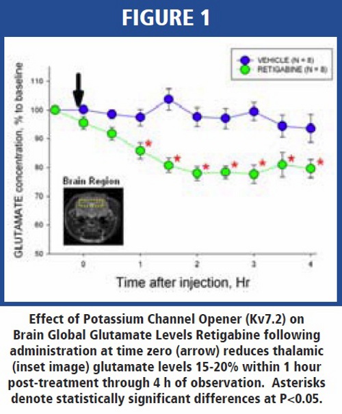
Mechanism based imaging biomarkers have been used to demonstrate drug action at the target through measurement of specific biological pathways. MRS through measurement of global brain pools of glutamine and glutamate has been used to link the effect of a Kv7 potassium channel opener with alterations in the glutaminergic pathway [3]. Increased cholinergic activity in vivo inhibits the M-current comprised of Kv7(2-5) potassium channel subunits, and thereby increase the resting membrane potential of neurons which reduces the excitation threshold required for generation of action potentials. At the synapse, the propagating action potential facilitates release of neurotransmitters such as glutamate. Retigabine, a Kv7 potassium channel opener, hyperpolarizes the neuron, reduces action potential and acting through the glutaminergic pathway decreases hippocampus glutamate levels by 20% which is detectable by MRS at 7 Tesla (Figure 1). Application of MRS to assessment of changes in the glutaminergic pathway is a sensitive measure to link drug action with a biological pathway; however, the local synaptic change must be associated with a global brain change in the specific neurotransmitter because MRI does not have the resolution to detect changes in individual synapses.
Positron emission tomography is a very sensitive molecular measure of changes in tumor metabolism as assessed through FDG but PET can also provide mechanistic information about specific drug targets based on the tracer being used. FLT-PET is a platform tool for measuring cell proliferation. Checkpoint kinase 1 (Chk1) is responsible for repair of DNA damage and inhibitors of the enzyme such as PF00477736 in combination with docetaxel have demonstrated efficacy by reducing tumor size through decreased cell proliferation which is detectable by FLT-PET [4]. FLT-PET can also be valuable in randomizing patients based on tumor proliferation rate prior to putting them on an anti-proliferative drug therapy. Preclinically, tumor bearing mice were randomized based on baseline level of tumor proliferation as assessed by FLT-PET [5]. In drug treated animals, docetaxil increased FLT uptake in tumors with low levels of proliferation (<1.6 max tumor SUV/mean liver SUV) at baseline but significantly decreased FLT uptake in tumors with a higher proliferation rate at baseline. Thus, these results have demonstrated that mechanism based biomarkers can allow one to characterize the drug effect on a specific mechanism like cell proliferation as well as allow one to use the mechanism to select the potentially most responsive patients.
While FLT and MRS were highlighted as examples of mechanism biomarkers, one can easily utilize FDG-PET to assess metabolic changes in tumors, degree of inflammation associated with rheumatoid arthritis [6], COPD, or atherosclerosis [7]. It is also possible to measure other mechanistic events like hypoxia or neuroinflammation based on the target tracer utilized, e.g., FMISO for hypoxia or PBR06 for neuroinflammation. The application of MRS while shown to be a sensitive marker for measuring glutamate changes can be expanded to measurement of other nuclei like C13 and C13-glucose or F19 to broaden the target species and/or disease area, e.g., C13-glucose and liver metabolism associated with diabetes.
Pharmacologic and Safety-Related Biomarkers
Upon establishing that a new pharmacologic agent is present in the target organ of interest and binds to the specific target, it is equally important to understand whether the compound has any pharmacological effect. Assessment of a pharmacologic effect does not imply efficacy, i.e., stabilization of atherosclerotic plaque or decreased osteoarthritis progression, but rather a measure of a drug action that may be implicated in a beneficial or safety outcome. AMPA receptor potentiators like PF00603173 are being explored as potential novel therapeutics for cognition enhancement. In rats, increases in FDG-measured glucose metabolism occurred in the cortex and thalamus at doses (0.125 and 0.25 mg/kg) associated with improved memory on cognition tasks (Figure 2) [8]. In contrast, a high dose (2.5 mg/kg) produced an increase in FDG uptake in cerebellum which was linked to cerebellum-mediated AEs (Figure 2). Thus, for AMPA receptor potentiators FDG-PET can be used as both a central measure of pharmacologic effect and a measure of safety which precedes motor related adverse side effects.
Such biomarkers of pharmacology or safety are not limited to FDG-PET. MRI has been utilized to demonstrate that a measure of extravascular water, i.e., edema, associated with acute exposure to calcium channel blockers (CCB) can be used to demonstrate that T-type CCBs may reduce vasodilatory edema related to L-type CCBs [9]. Measures of cerebral blood flow, ASL-MRI, have also been developed as a platform tool to assess changes related to Alzheimer’s and evaluated in transgenic Tg2576 mice as an early marker for assessment of drugs noted to improve cognition [10]. Finally, general measures of brain activation as assessed by a functional MRI technique known as BOLD MRI is a sensitive measure of brain oxygen utilization following drug or pain stimuli like electric forepaw stimulation that is not only stimulus strength dependent but also time dependent [11].
While the power of the various types of platform imaging biomarkers lies in the ability to study specific processes with a common tool across different diseases, greater benefit might be achieved by combining the tools to better understand the disease process. It is tempting to speculate that imaging biomarkers can be utilized to link target to clinical efficacy. One might propose that imaging can be used to demonstrate the following sequence of biologic events: drug binding to a receptor associated with Alzheimer’s progression which modifies amyloid burden resulting in increased cerebral blood flow, oxygen utilization, neuronal metabolism, synaptic activity and improved cognition. The imaging tools that can be applied to assess the above events can be represented by the following scheme: Target PET Tracer -Amyloid PET Tracer -ASL MRI -BOLD MRI -FDG PET -MRS -Clinical Memory Test.
Summary and Future Horizons
Platform imaging biomarkers focus on mechanistic changes and have the potential to be used across multiple disease applications. These platform tools can provide a means of assessing drug target organ exposure, target binding, proof of mechanism and pharmacologic/safety effect of a new drug. The biomarkers allow for translation of findings from preclinical studies where histology or an invasive procedure may be the efficacy endpoint to the clinic and in some cases biomarkers with utility in the clinic can be back translated to animals to better qualify the various animal models. With advancement in molecular imaging tools, imaging can provide a means to advance our understanding of disease mechanisms and allow one to assess viability of the target, mechanism and pharmacologic agent to potentially reduce compound attrition at later stages of development. Through broadening the use of currently available imaging tools and methods, building novel small molecule or protein-based PET tracers and targeted MR and CT contrast agents, imaging biomarkers can potentially be used to study novel biologic agents such as proteins, antibodies and antibody fragments, siRNA and stem cells and provide greater opportunities for studying conventional pharmacologic agents.


References
1. Paul SM, Mytelka DS, Dunwiddie CT, Persinger CC, Munos BH, Lindborg SR, Schacht AL. How to improve R&D productivity: the pharmaceutical industry’s grand challenge. Nat Rev 2010 9:203-214.
2. Zasadny K, Callahan MJ, Watson MF, Li Z. Comparison of dopamine D2 receptor occupancy measurements by ex vivo binding assay versus in vivo microPET imaging for typical and atypical antipsychotics (meeting abstract). J Nucl Med 2006 47(5): 9P
3. Sriram T, Mather RJ, Harris RL, Burkholder A, and Liachenko S. Acute Retigabine Administration Reduces Level of Glutamate in Rat Hippocampus ISMRM 2009.
4. Cathy Z, Yan Z, Painter CL, Zhang Q, Chen E, Arango ME, Kuszpit K, Zasadny K, Hallin M, Hallin J, Wong A, Buckman D, Sun G, Qiu M, Anderes K, Christensen JG. PF- 00477736Mediates Checkpoint Kinase 1Signaling Pathway and Potentiates Docetaxel-Induced Efficacy in Xenografts Clin Cancer Res 2009 15(14):4630-4640.
5. Kuszpit K, Callahan M, Zhu A, Burkholder A, Harris R, Chen L, Zasadny K [18F]-FLT PET of mouse xenograft tumor models for assessing treatment response: Making a case to randomize treatment groups based on baseline tumor FLT uptake and not tumor volume WMIC 2009.
6. Callahan MJ, Zasadny KR, Chen L, Lesch M. FDG PET imaging assessment of inflammation in a rheumatoid arthritis model in the rat. AMI 2006.
7. Zasadny KR, Callahan MJ, Chen L, Liachenko S. FDG PET imaging in a rabbit model of atherosclerosis: Comparison of uptake in two models aimed at producing characteristics of unstable versus stable aortic plaques. WMIC 2009.
8. Callahan MJ, Kuszpit K, Chen L, Skaddan MB, Brown-Proctor C, Harris R, Zhu A, Shaffer C, Scialis R, Zasadny KR. 2-Deoxy-2- [F-18]flouro-D-glucose Positron Emission Tomography Imaging Provides Insights into Efficacy and Safety for an AMPA Receptor Potentiator WMIC 2009.
9. Major TC, Dhamija S, Black N, Liachenko S, Morenko B, Sobocinski G, Okerberg C, Tinholt P, Madore S, Kowala MC. The T- and L-Type Calcium Channel Blocker (CCB) Mibefradil Attenuates Leg Edema Induced by the L-Type CCB Nifedipinein the Spontaneously Hypertensive Rat: A Novel Differentiating Assay. J Pharm Expt Therap 2008 325(3):723-731.
10. Goodman JA, Xie Z. Stability of Repeat Measures of CBF in Aged Tg2576 and Wild Type Mice via CASL. ISMRM 2010. 11. Yang D, Xie Z, Goodman J, Burkholder A, Poy N. Temporal hemodynamic responses of BOLD fMRI in the rat brain related to electric forepaw stimulation. ISMRM 2010.
Dr. Thomas Bocan is the Senior Director and Head of Pfizer’s PharmaTherapeutics preclinical BioImaging facility. Dr. Bocan received his PhD in Physiology with an emphasis in Experimental Pathology from Penn State University in 1983 and did post-doctoral research in atherosclerosis at the Baylor College of Medicine. In 1986, he joined the Atherosclerosis Pharmacology Department at Parke-Davis as a Senior Scientist. Dr. Bocan was awarded the Warner- Lambert 1991 Meritorious Scientific Achievement Award for his research efforts in atherosclerosis. Dr. Bocan has chaired or co-chaired numerous drug discovery teams during his tenure in Cardiovascular Pharmacology. He has co-authored the preclinical Pharmacology summaries for both the atorvastatin IND and NDA as well as for the clinical candidate ACAT inhibitor, CI-976. Tom has been a member of several drug development teams and was the preclinical expert on the Lipitor development team from 1998 to 2001. In July 2001, Tom became Director of Pfizer’s Preclinical BioImaging Facility and assumed responsibility for building and managing the facility. Dr. Bocan has authored 50 manuscripts, 65 abstracts, 44 internal research reports and is the inventor on 6 patents. He has trained 2 post-doctoral fellows and managed a staff of 12-15 scientists.