Abstract
This application note reports the development of two rapidly reacting fluorescent dyes (Teal™ and Turquoise™ dyes) for N-glycan analysis. The dyes also eliminate the use of toxic sodium cyanoborohydride from the labeling chemistry. A number of N-linked oligosaccharides (IgG glycans) consisting of both charged and neutral glycans, some of which were unresolved when labeled with conventional aminopyrene trisulfonic acid (APTS) dye, were fully baseline-resolved when labeled with our proprietary dyes. N-glycans labeled with these novel dyes can be analyzed using a liquid chromatography (LC) or capillary electrophoresis (CE) separation platform. These dyelabeled N-glycans can also generate better signals when analyzed using mass spectrometry methods.
Introduction
Glycosylation is one of the key critical quality attributes of monoclonal antibody–based biotherapeutics [1,2]. Glycosylation changes can impact a biological drug’s safety, efficacy, clearance, and immunogenicity, making it necessary to accurately detect changes. Glycan profiling begins at cell line development, continues through process development, and in certain cases continues through drug substance release. Current glycan analysis methods involve laborious multistep sample preparation that takes anywhere from a day to multiple days for 96 samples, followed by single-channel LC or CE separation. Here we report an integrated glycan profiling solution that can generate data from 96 samples in 7–9 hours, consisting of an easy magnetic bead–based sample preparation, 24-capillary array CE instrument, and a glycan-specific software for analysis (Figure 1).

Figure 1. The Applied Biosystems™ GlycanAssure™ glycan analysis workflow. Only 7–9 hours (with 3 hours of hands-on time) are required to process and analyze 96 samples, with no vacuum centrifugation steps or use of sodium cyanoborohydride.
Materials and Methods
All CE separations were performed using the Applied Biosystems™ 3500xL Genetic Analyzer (Cat. No. A30887), a system configured with a 505 nm solid-state laser and laser-induced fluorescence detection. Glycans were separated using 24-capillary arrays with Applied Biosystems™ POP-7™ Polymer (Cat. No. A30936) and cathode and anode buffers (Cat. No. A31279, A31278). Additional experimental details for CE analysis were as follows:
- Capillary length: total length = 61 cm, length to detector = 50 cm
- Capillary diameter: 50 μm ID
- Injection conditions: 1.6 kV for 24 sec
- Run voltage: 19.5 kV
- Capillary oven temperature: 60°C
- APTS: 475 nm (Ex), 501 nm (Em)
- Teal dye: 466 nm (Ex), 505 nm (Em)
- Turquoise dye: 493 nm (Ex), 520 nm (Em)
All other assay conditions were as described in the N-Linked Glycan Analysis User Guide (Pub. No. 100033998). Invitrogen™ Purified Human IgG (Cat. No. 027102) and glycan standards from ProZyme and V-Labs, Inc., were used for analysis.
Results and Discussion
The goal of the project was to develop novel fluorescent dyes to label glycans and combine them with a highthroughput separation method to quantitate complex glycan species associated with therapeutic glycoproteins. These glycans include structures with and without a core fucose moiety, terminal galactose, terminal sialic acids, high- mannose structures, and several positional isomers. Cleaved glycans were purified using magnetic beads and labeled with fluorescent APTS, Teal dye, and Turquoise dye using optimized labeling conditions. Excess dye was removed using the same magnetic beads used for glycan purification. CE separation was performed using optimized conditions. Separation efficiencies for glycans labeled with the novel fluorescent dyes were benchmarked against those for glycans labeled with the widely used APTS. For glycans labeled with APTS, we observed co-migration of A1F and Man5 glycans. These glycans were baseline-resolved when labeled with Teal or Turquoise dye (Figure 2). We attained the best resolution with Teal dye, where not only A1F/ Man5 and G2F/Man9 glycan pairs, but also positional isomers of A1, Man6, G1F, Man8, and Man9, were resolved (data not shown).
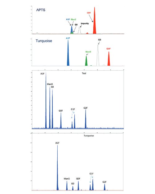
Figure 2. Separation of major glycans with APTS, Teal, and Turquoise dyes. APTS did not resolve A1F and Man5 glycans. Glycans not resolved with APTS can be resolved with novel Teal and Turquoise dyes.
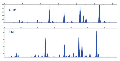
Figure 3. Separation of labeled N-glycans from human serum IgG.Teal dye resolves glycans better than APTS does.
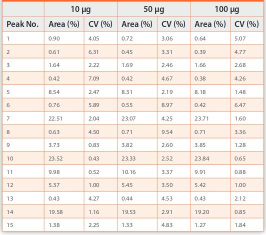
Teal dye resolves N-glycans from human serum IgG better than APTS does (Figure 3). Consistent relative quantities of these glycans were obtained from varying inputs of human serum IgG in the range of 10−100 μg (Table 1). Eight independent sample preparations were performed for every input amount. Higher variation was observed in peaks with a relative percent area lower than 1%.
Table 1. Consistent relative quantities of glycans from varying inputs of IgG.
Normalization with internal size standards co-migrating in every capillary revealed highly reproducible separation of multiple glycan species (12 injections across 24 capillaries, 288 total injections; Figure 4).
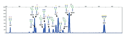
Figure 4. Overlay of 288 injections from 24 capillaries on the 3500xL Genetic Analyzer. Glycan standards were labeled with APTS.
Conclusions
The GlycanAssure workflow offers the following benefits for N-glycan analysis:
- Streamlined workflow—new dyes, software, and automatable purification enable parallel processing of 96 samples with analysis on the 3500xL Genetic Analyzer in less than 9 hours
- Resolution—Teal and Turquoise dyes offer better resolution of key glycans
- Precision—high run-to-run, capillary-to-capillary, and instrument-to-instrument reproducibility (data not shown) supports high-throughput analysis of protein glycans
References
- Varki A (1993) Biological roles of oligosaccharides: all of the theories are correct. Glycobiology 3:97–130.
- Bertozzi CR, Freeze HH, Varki A, Esko JD (2009) Chapter 51: Glycans in biotechnology and the pharmaceutical industry. In: Essentials of Glycobiology, 2nd edition. Cold Spring Harbor (NY): Cold Spring Harbor Laboratory Press.