Introduction
Polyethylene glycols (α-Hydroxy-ω-hydroxy-poly(oxy-1,2ethanediyl) abbreviated PEGs, are a family of non-ionic synthetic polymers with an empirical formula HOCH2(CH2OCH2)mCH2OH (1). The subscript m is a measure of the average number of oxyethylene units present in the polymer chain and forms the basis of classification of PEGs. Physical properties of pure PEGs and of aqueous solutions of PEGs have been extensively studied and reported (1, 2). It is observed that an increase in the molecular weight of PEG results in an increase in the density and viscosity of the solution. Table 1 lists some of the key PEGs and the associated properties. Prior knowledge of the physical properties of PEGs may be of significance at the time of selection of a PEG for its potential applicability.
Table 1.

These randomly coiled non-ionic polymers have been extensively used for the crystallization and precipitation of proteins for various purposes (3-5). They are used both in academia and industry for the purification of proteins from blood plasma, precipitation of proteins for the crystallographic studies and for the estimation of protein activities in saturated solutions (5-7). PEGs have been shown to separate free hormones from those bound to membranes and/or receptors and free antigens from antibodies, leading to the development of improved immuno- and bio-assays (8-10). Recently, Sharma and Kalonia have reported that precipitation of proteins by PEGs under favorable conditions followed by vacuum drying of the precipitate can be used for the formulation of proteins in the dried form (11). Use of these synthetic non-ionic polymers for the purpose of protein precipitation was first introduced by Polson et al. in 1964 (12). Since then, there has been tremendous gain in the knowledge of the precipitating action of these polymers. The reason in part being the growth in the development of biotechnology based drug therapeutics. The developments have provided improved understanding of the effect of solution conditions on the PEG induced precipitation, impact of PEGs on the secondary and/or tertiary structure of proteins, molecular basis of the precipitating action, and the advantages offered by PEGs over other commonly used precipitating agents. The purpose of the present chapter is to review and summarize these aspects such as to enable the readers to assess the potential applicability of the polymers for their specific usage, especially during the early stage formulation development.
Mechanism
Precipitation work on a wide variety of purified proteins shows that the dependence of protein solubility, S, on PEG concentration is strictly exponential and that the data usually adhere to the following equation (13):
log S = log So – βC (1)
Where, C is the polymer concentration, S is protein solubility in the presence of PEG at concentration C (% w/v) and So is the apparent solubility at zero polymer concentration obtained from extrapolation. The form of equation is identical to that used to describe protein solubility in the presence of salts (14). Figure 1 shows the effect of several PEGs on the solubility of human serum albumin and exemplifies the linear relationship that is obtained when PEG concentration is plotted against log of protein solubility (13).
Thermodynamic analysis of the protein-polymer system led authors to arrive at an equation that substantiates the observed dependence of protein solubility on PEG concentration. This equation, has been derived based on the chemical potential of the protein under conditions of saturation in the absence and presence of PEGs (13).
In S3 = ln S’3 + dS’3 – aC2 (2)
Where, S3 and S’3 are protein solubilities in the absence and presence of PEG, d and a represent protein-protein and PEG-protein interactions, respectively and C2 is the concentration of PEG. When solubility is expressed in grams/liter and PEG concentration is expressed in % w/v, a and d can be obtained from the following expressions,
a = -0.23ΒM2
d = (2.3M3)/(S’3) log (S’3 apparent)/(S’3) = (2.3 M3)/(S’3) log γ
Where β is the measure of the slope of precipitation curve, M2 is the average molecular weight of PEG, M3 is the molecular weight of the protein, S’3 (apparent) is obtained from extrapolated intercept and γ is the thermodynamic activity coefficient of the protein under saturating conditions in the absence of PEG.

Figure 1 - Effect of molecular weight of PEG on the solubility of human serum albumin. Measurements were made with a total protein concentration of 20 mg/ml at pH 4.5 buffer containing 0.1 M KCl (data re-plotted from reference 13).
Figure 1 also shows that the effectiveness of PEG in reducing the solubility of protein increases with increasing size of the polymer (slopes increase with increase in the size of PEG). It is clear that within the experimental error, the intercepts are constant, indicating that neither d nor S’3 depend on the type of PEG. The slopes of log S vs. polymer concentration in general also tend to increase with an increase in the size of the protein used. These results along with the results of the effect of solution conditions on the precipitation of the proteins suggest the absence of any chemical interactions between proteins and PEGs. The dependence of macromolecular size on the effectiveness of precipitation when combined with the lack of any specific chemical interactions seems compatible with the notion of steric exclusion mechanism of protein precipitation. The phenomenon of steric exclusion results in preferential interactions that depend only on the number, size and shape of the molecules involved (15). In the present case, the effect arises because of the difference in the size of the solvent and co-solvent molecules. Because of the larger size of the co-solvent molecules as compared to the solvent molecules, there is a region surrounding each protein molecule from which the co-solvent is excluded but the solvent can enter. As a result, there is a concentration gradient between the co-solvent rich bulk phase and co-solvent depleted local domain surrounding the protein molecules. Presence of co-solvents thus results in an increase in the chemical potential of protein because of the greater free energy required to maintain this concentration gradient.
Support for the steric exclusion theory came from some pioneering work done by Timasheff and colleagues (16-18). These authors utilized dialysis equilibrium for the measurement of the preferential interactions in water/protein/PEG ternary systems. Several significant observations were made from the results that were obtained. Values of dg1/dg2 (preferential hydration parameter) were found to be positive for all combinations of PEGs and proteins that were studied, indicating that PEGs are preferentially excluded from protein domain (17). Furthermore, the magnitude of preferential hydration was found to increase with an increase in the size of PEG. They also utilized the preferential interaction parameters for calculating the effect of PEGs on the chemical potential of the proteins and found that an increase in the size of PEG does indeed result in an increased effect on the chemical potential of the protein (17).
Despite the fact that most literature data supports the role of steric exclusion, the extent of the role remains questionable because of the strong discrepancies that have been observed in this regard. Simple theoretical calculations based on excluded volume considerations by Donald et al. have shown that the predicted influence of PEG size on protein precipitation does not match with the experimental observations (13). Attempts have also been made to theoretically predict protein solubilities in the presence of varying molecular weight PEGs (19). Though, several of these attempts have been able to predict the trends that are observed, none of the theories have so far been able to predict the experimental solubilities to an acceptable degree. Similarly, the predicted influence of protein size on the slope of the precipitation curve is also at variance with the experimental values. Furthermore, Mahadevan et al. have shown that linearity of the protein solubility/PEG concentration curve does not extrapolate to zero polymer concentration (19).
Structural studies done towards the understanding of specific interactions between PEGs and proteins have given insight into the mechanism of protein precipitation. Furthermore, the knowledge of the effect of PEGs on the structural stability of proteins is important when proteins under study are being precipitated for their therapeutic applications. Authors have utilized a variety of spectroscopic (Circular Dichroism, differential UV absorption etc.) and calorimetric techniques in order to determine any such specific interactions that might occur between PEGs and proteins (4, 11, 20-23). Results from the spectroscopic measurements have shown that PEGs in general do not perturb the native structure of proteins. Lee et al. studied the effect of 10 % w/v PEG 1000 on the far and near UV-CD spectra of lysozyme and concluded that presence of PEG does not significantly alter the native structure of the protein (16). Donald et al. investigated the effect of 10% w/v PEG 4000 on the secondary and tertiary structure of ribonuclease and came up with similar conclusions (13). Although, spectroscopic studies show that PEGs have little effect on protein structure, thermal denaturation studies have shown that most PEGs decrease the thermal unfolding temperature of proteins in solution. Lee et al. showed that the magnitude of destabilization depends on both, the type of protein and the type of PEG, and that the extent of destabilization increases with an increase in the concentration of PEG (16). Furthermore, the extent of destabilization was also found to be highly dependent on the hydrophobicity of the protein. Farruggia et al. investigated the effect of different molecular weight PEGs on the thermal stability of human serum albumin and observed that a decrease in the molecular weight of PEG decreased the thermodynamic stability of the protein (20). In fact, some of the higher molecular weight PEGs (> PEG 8000) were found to increase the unfolding temperature, albeit to a very small extent. The observations are consistent with the fact that PEGs have weak hydrophobic nature and hence can interact favorably with the non-polar patches of the protein. Considering that proteins fold due to hydrophobic interactions, enhanced propensity of proteins to unfold in the presence of hydrophobic PEGs seems a reasonable observation.
Although, it is clear that PEGs can interact with proteins and caution must be exercised in the use of these additives especially under extreme conditions such as high temperatures, the role of specific interactions in the process of precipitation is not yet well understood. Considering that little phase separation of PEG occurs during the process of protein precipitation, it seems reasonable to assume that such PEG-protein interactions should in fact result in an increase in the solubility of the protein. Therefore, it seems quite possible that favorable interactions between PEG and protein can counteract the effect of steric exclusion of PEG from protein domain.
Formulation Variables
Knowledge of the effect of solution conditions on the precipitation of proteins by PEGs not only provides insight into the mechanism but is also helpful during the design of experiments for particular specific usage of PEGs. In general it has been observed that the amount of PEG required to precipitate the protein out of the solution is highly dependent on the formulation conditions (such as pH, ionic strength, temperature etc.) used for the study (4, 11). The reason in most part being the fact that protein solubility by itself (in the absence of PEGs) is significantly dependent on the solution conditions. For compact protein ions, solubility has been described by the following relationship (13)
ln (S2/S2,w) = (Z2εNκ)/[2DRT(1 + κa)] (3)
Where Z is the net charge on the protein, ε is the electronic charge, N is Avogadro’s number, D is the dielectric constant of the medium, T is the temperature, R is the gas constant, a is the sum of the radii of the protein ion and the supporting electrolyte ions in solution and κ is given by
κ = [(8ΠNε2)/(1000DkT)]1/2(I)1/2
I = ½ (ΣCiZi2)
Where I is the ionic strength and k is the Boltzmann’s constant. As can be observed from the above equation, protein solubility is expected to be significantly affected by factors such as the ionic strength, pH and the polarity of the solvent.
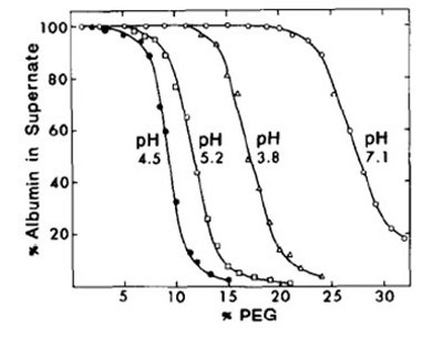
Figure 2 - Effect of pH on the precipitation of albumin by PEG 4000 at room temperature. All solutions contained 0.1 M KCl and the concentration of albumin was 20 mg/ml (reference 4).
An example of the effect of pH on the solubility of proteins in the presence of PEGs is demonstrated in figure 2 which shows pH/solubility profiles of bovine and human serum albumin in the presence of PEG-4000 (11) (it should be realized that the log S verses % PEG is not linear in this case). The protein was observed to have lowest solubility at around its reported isoelectric point (pI = 4.60) (13). Since, proteins have no net charge around the pI (Z = 0), protein-protein repulsive interactions (and hence protein solubilities) are anticipated to be minimum at the isoelectric point.
The above equation also shows that at the isoelectric point (Z=0), protein solubility should increase with an increase in the ionic strength of the solution. Addition of salt screens the charges on the protein. This screening of charges decreases the free energy of the protein resulting in a decrease in its activity and increase in its solubility. Hence, at low concentrations and close to the isoelectric point of the protein, salts usually have a salting-in effect on protein solubility (14, 24). On the other hand, at high salt concentrations and far from the isoelectric, a salting-out effect is observed, which occurs due to the increase in the activity coefficient of the protein. Sharma and Kalonia studied the effect of solution ionic strength on the apparent solubility of IFNα2a at 25°C and pH 6.5 (6.5 is the pI of the protein) (11). A decrease in the ionic strength significantly decreased the solubility of IFNα2a. The effect of salts on the precipitation of albumin also correlates well with the position of the salts in the Hofmeister or lyotropic series (series which rank salts in the order of their effectiveness to precipitate colloids). Evidently, the data present in literature show that effect of solution conditions on the solubility of proteins in the presence of PEGs is similar to the effect that are anticipated to be observed in the absence of PEGs. The measurement of protein solubility in the presence of PEG thus provides an easy and alternative way to obtain information about the relative solubility of the protein under different solution conditions.
One potential advantage of using PEGs for the precipitation of proteins lies in the fact that rigorous control of temperature is not required. Sharma and Kalonia investigated the effect of temperature (5°, 25° and 37°C) on the apparent solubility of IFNα2a in the presence of varying concentrations of PEG 1450 and concluded that temperature has no significant effect on the solubility of the protein (11). Similar results have also been obtained by other authors (4).
Salts, Organic Solvents and PEGs: A Comparison
A lot of precipitants including salts (Na2SO4) and organic solvents (ethanol) are available that can be used to phase separate proteins out of the solution (14). For processes such as purification and fractionation, wherein there is a step involved for the removal of precipitant, any of the commonly available precipitating agents can be used. However, if the precipitate is to be used for therapeutic applications and/or the precipitating agent is anticipated to form the integral part of the therapeutic formulation, extreme care is desired during the selection of the precipitating agent. In such cases it becomes important that the desired precipitating agent have all or some of the following properties:
- It should not perturb the native structure of the protein (and hence compromise protein’s activity) and that the protein should preserve its activity on reconstitution.
- The precipitant should be effective in small concentrations.
- The precipitant should be approved for parenteral use.
Organic solvents such as ethanol, though effective are required in high concentrations. It is also well established in literature that organic solvents perturb the native structure of the protein molecules. Even small concentrations of these solvents have been shown to significantly decrease the thermal denaturation temperature of proteins. Zaks et al. investigated the effect of organic solvents on the catalytic activity of several enzymes and showed that the activity of most proteins is severely compromised in non-aqueous solvents (25). Additionally most organic solvents are not approved for parenteral use and their presence even in small concentrations may result in unwanted toxic effects. Similar issues as observed for organic solvents are true for salts. Most salts such as ammonium sulfate are required in high concentrations, and high concentration of salts have been shown to decrease the thermodynamic stability of proteins in solution (14, 26). PEGs offer another advantage over salts and organic solvents. PEGs have been shown to facilitate the growth of protein crystals which in turn reduces the time required to achieve complete crystallization. In a nutshell, PEGs satisfy most of the criterion of perfect precipitating agents and hence can conveniently be used as the agents of choice for a wide variety of purposes wherein protein precipitation is desired.
Summary
Polyethylene glycols are water soluble synthetic polymers whose ability to precipitate proteins out of the solution can most convincingly be understood in terms of the concept of excluded volume. Though, extreme conditions such as high temperature may result in unwanted favorable interactions between PEGs and proteins, PEGs under normal conditions do not perturb the native structure of the protein molecules. PEGs offer several advantages over some of the other commonly available precipitating agents and fulfill most criterions of perfect precipitating and crystallizing agent. The technique of PEG assisted solubility estimations and/or comparisons perhaps offer greatest advantage to the preformulation scientists. Candidate screening during early development could easily be done in a 96 well plate format wherein the plate could be read on a UV plate reader following precipitation.
References
- B. S. Windholz M, Stroumtsos LY, Fertig MN, Ed., Polyethylene glycol (monographs), (Merck and Co Inc., Whitehouse Station, NJ, 1976), pp. 983-984.
- . P. JC, Ed., Polyethylene Glycol, (The American Pharmaceutical Association, Washington DC, 1994), pp. 355-361.
- R. N. Haire, W. A. Tisel, J. G. White, A. Rosenberg, Biopolymers 23, 2761 (1984).
- K. C. Ingham, Arch. Biochem. Biophys. 186, 106 (1978).
- P. H. Iverius, T. C. Laurent, Biochim. Biophys. Acta, Protein Struct. 133, 371 (1967).
- P. W. Chun, M. Fried, E. F. Ellis, Anal. Biochem. 19, 481 (1967).
- C. R. Middaugh, W. A. Tisel, R. N. Haire, A. Rosenberg, J. Biol. Chem. 254, 367 (1979).
- W. D. Creighton, P. H. Lambert, P. A. Miescher, J. Immunol. 111, 1219 (1973).
- P. Cuatrecasas, Proc. Nat. Acad. Sci. U. S. 69, 318 (1972).
- M. L. Dufau, E. H. Charreau, K. J. Catt, J. Biol. Chem. 248, 6973 (1973).
- V. K. Sharma, D. S. Kalonia, AAPS PharmSci 6, E4 (2004).
- A. Polson, G. M. Potgieter, J. F. Largier, G. E. F. Mears, F. J. Joubert, Biochim. Biophys. Acta, Gen. Subj. 82, 463 (1964).
- D. H. Atha, K. C. Ingham, J. Biol. Chem. 256, 12108 (1981).
- T. Arakawa, S. N. Timasheff, Methods Enzymol. 114, 49 (1985).
- D. J. McClements, Crit. Rev. Food Sci. Nutr. 42, 417 (2002).
- L. L. Y. Lee, J. C. Lee, Biochemistry 26, 7813 (1987).
- R. Bhat, S. N. Timasheff, Protein Sci. 1, 1133 (1992).
- T. Arakawa, S. N. Timasheff, Biochemistry 24, 6756 (1985).
- H. Mahadevan, C. K. Hall, AIChE J. 36, 1517 (1990).
- B. Farruggia, G. Garcia, C. D’Angelo, G. Pico, Int. J. Biol. Macromol. 20, 43 (1997).
- B. Farruggia, B. Nerli, N. H. Di, R. Rigatusso, G. Pico, Int. J. Biol. Macromol. 26, 23 (1999).
- W. Zielenkiewicz, R. Swierzewski, F. Attanasio, G. Rialdi, J. Therm. Anal. Calorim. 83, 587 (2006).
- K. Gekko, J. Biochem. 91, 1197 (1982).
- D. Forciniti, C. K. Hall, M. R. Kula, Biotechnol. Bioeng. 38, 986 (1991).
- A. Zaks, A. M. Klibanov, J. Biol. Chem. 263, 3194 (1988).
- A. Saluja, A. V. Badkar, D. L. Zeng, D. S. Kalonia, J. Pharm. Sci. 96, 3181 (2007).
Author Biography
Vineet Kumar, Ph.D. is a Sr. Scientist at Abbott. He earned his MS (2002) and Ph.D.(2007) in Pharmaceutical Sciences from University of Connecticut. In his current role he is primarily focused on preformulation characterization and early stage formulation development of therapeutic biological drug candidates. He has been invited to present his work at several conferences and has several publications, patents and abstract publications to his credit.
Nitin Dixit is a Ph.D. candidate at the University of Connecticut.
Shubahdra N. Singh, Ph.D. is a postdoctoral fellow at the University of Connecticut.
Professor Devendra S. Kalonia, currently at the University of Connecticut, is a known expert in the area of protein formulations and delivery.
This article was printed in the November/December 2011 issue of American Pharmaceutical Review - Volume 14, Issue 7. Copyright rests with the publisher. For more information about American Pharmaceutical Review and to read similar articles, visit www.americanpharmaceuticalreview.com and subscribe for free.