Abstract
The performance of functional foods and pharmaceutical products is highly dependent on nano-scale self assembly of lipids and other substances in the gastrointestinal tract. Digestion of the lipids in lipid-based formulations produces complex structures in the gastrointestinal tract that act to maintain drug in solution and hence improve consistency in absorption and bioavailability. However, despite these structures being critical to the outcome, little is known about their structure at the ‘nanoscale’ during digestion and hence limits our ability to rationally select the best lipids for the job. The research described in this article seeks to use contemporary advanced techniques, in particular synchrotron small-angle X-ray scattering, in order to understand the structural changes that occur during the digestion of lipids to ultimately enable the link between composition, structure and performance to be established.
Lipids are common molecules found in cell membranes, fats, oils and plant tissue. It is well known that amphiphilic lipids (lipids that possess molecular features that have opposite affinity for water and oil) will self-assemble in water and biological fluids to form structured systems such as liquid crystalline phases, vesicles and micelles (right hand panel Figure 1). Nature uses this phenomenon for a number of biological purposes, including the formation of bilayer membranes to provide cellular structural integrity, and to form nano-carriers for non-polar compounds in the bloodstream and the lymphatic system, and through lipid digestion in the gut. The manner in which they self-assemble depends on composition and conditions such as temperature and pressure, as well as the molecular geometry of the amphiphilic compounds present. Subtle changes in any of these variables can lead to a dramatic change in the self-assembled structure. Despite the promise of lipid-based formulations to improve the bioavailability of poorly water soluble, highly permeable (BCS Class 2) compounds, until the link between structure, composition and outcome are established formulation approaches will remain largely empirical.
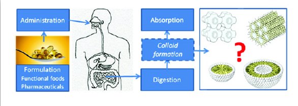
Figure 1 - Self assembly of lipid-based colloids in the gut to form nanoassemblies is the determining factor in subsequent absorption processes – only by fully understanding this step can we hope to design new highly eff ective function
Approaches to Understand Colloid Formation in the Gut, Even In-vitro, have not Progressed since the 1970s
The self assembly of lipids in the gut has been recognized for decades, since the observation of a micellar phase in the intestine in 1962 [1]. The seminal work of Patton and Carey “Watching Lipid Digestion” [2] in 1979, was the first direct observation of liquid crystalline structure formation on exposure of lipids to gastrointestinal fluids. Separation of the digesting oil, liquid micellar and solid pellet phases by centrifugation, and subsequent characterization of lipid content, was undertaken to enable conclusions to be drawn on the basis of composition. Accepted techniques for studying lipid digestion have not progressed beyond this simple approach. This statement is most evident from a recent review in Nature Reviews – Drug Discovery on lipids and lipid digestion, which provides an in-depth description of the current techniques applied to study lipid digestion where it is clear that this approach is still state of the art [3].
The Patton and Carey “in vitro lipolysis model” has subsequently been used now for four decades, but it is apparent that our understanding of lipid processing in vivo has not progressed substantially beyond that described in 1979. Contention over the composition of simulated gastrointestinal media, and methods of application of the ‘lipolysis model’ approach, have limited both the value of information obtained from the model, as well as inhibited progression onto more valuable techniques – the lipolysis models do not provide any direct dynamic structural information.
Nano-structure Formation is a Critical Determinant in Absorption
We know that formation of liquid crystalline and lipid colloidal structures directly impacts on drug absorption [4]. However, currently there is a poor understanding of how composition in foods and pharmaceutical formulations impacts on nano-structure formation in the gut. Until direct structural understanding of nano-assembly in the gut is fully understood the current intense research in drug delivery and functional foods cannot make rational, step-change progress.
Nano-assembly in the Gut is a Dynamic, Non-equilibrium Process
Self assembled structure is often highly dynamic in real systems, and continual changes in composition and conditions can make the understanding of self assembled behavior difficult to interpret from static equilibrium information. The gastrointestinal tract is a highly complex lipid processing system. Regional changes in degree of mixing, pH, ionic strength and composition all occur simultaneously. It is apparent that any attempt to understand behaviour in such a complex system using static equilibrium approaches is unlikely to provide understanding of value that can be applied to ‘real’ systems.
Attempts to link composition with structure have been reported using small angle x-ray scattering (SAXS) and cryo-transmission electron microscopy (cryo-TEM). However, these studies have not been in real time, dynamic settings. Borne et al. have reported on structural evolution in monoolein and diolein based lipid systems when exposed to lipolytic enzymes using these techniques [5,6]. Structural evolution was correlated with equilibrium phase behavior – however dynamics in these systems are extremely important. More recently, Fatouros et al. have attempted to couple the in vitro lipolysis model to SAXS [7] and cryo-TEM [8] to attempt to understand the structural evolution occurring in digesting systems. While this approach is laudable and is a good step towards achieving understanding of the dynamic link between lipid digestion and structure in vitro, the approach required sampling of the digestion media, inhibition of lipolysis with additives, and significant delay before sample analysis, leading to uncertainty of representativeness of the sample at time of analysis. The approach still does not provide a real time, dynamic understanding of the lipolysis process from a structural perspective.
Application of Contemporary Advanced Methods
Hence there is a need to utilize advanced methods to understand non-equilibrium lipid behaviour and self assembly in vitro and in vivo. To this end, we have taken advantage of the high intensity and fast acquisition time of synchrotron radiation to provide real-time kinetic information of the structural evolution occurring during lipid digestion. The in vitro lipolysis apparatus [3] (see schematic in Figure 2) was equipped with a flow-through capillary cell, which was placed in the beam at the SAXS-WAXS beamline at the Australian Synchrotron. Briefly, lipid formulations were dispersed by stirring in 10 mL simulated intestinal fluid containing 5 mM sodium taurodeoxycholate and 1.25 mM phospholipid. The formulation was circulated through a glass capillary to enable in-line real-time scattering acquisition. Enzyme was added using a remotely operated syringe driver, and fatty acids generated were titrated using a pH stat to maintain pH 7.5 to enable digestion kinetics to be followed as described previously [3].
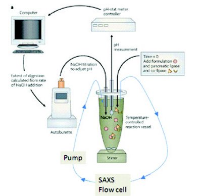
Figure 2 - In vitro digestion/SAXS schematic
Nanostructure in ‘Known’ Structured Systems
To establish the methodology, we conducted initial experiments using so-called ‘cubosomes’ – liquid crystalline particles with known internal structure. Two types of cubosomes were studied, one where the lipid (phytantriol, Figure 3) is not subject to enzymatic digestion, and hence the structure was expected to be retained over time, and the second, using glyceryl monooleate (Figure 4) which is subject to digestion and hence change in structure over time was anticipated. The characteristic peaks for the internal structure of cubosomes prepared using phytantriol did not change over time during digestion (Figure 3), although there was a slight shift in position when exposed to the bile salt medium required to facilitate digestion. In contrast, the digestion of glyceryl monooleate cubosomes induced dramatic shifts in peak positions (Figure 4) with eventual destruction of the internal order of the cubosomes over approximately 30 min (a physiologically relevant time scale)
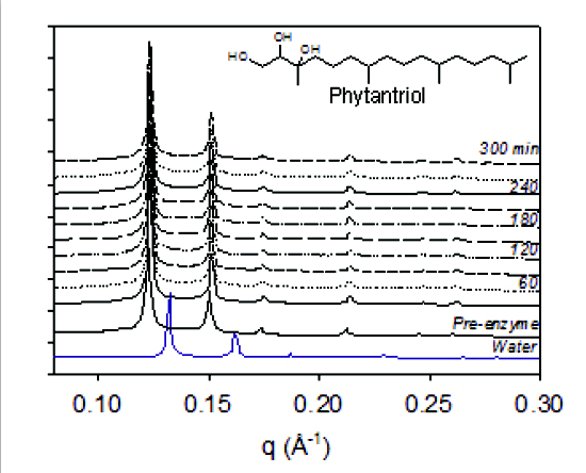
Figure 3 - SAXS profi les during digestion of phytantriol cubosomes
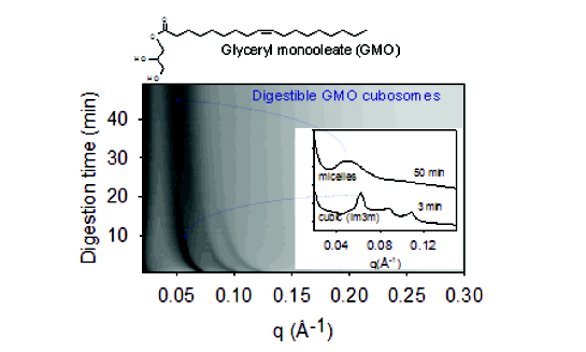
Figure 4 -SAXS profi les during digestion of glyceryl monooleate cubosomes
Nanostructure Evolution during Digestion of Unstructured Lipid Systems
Structure evolution during the digestion of a dispersed non- structured emulsion, containing common formulation lipids (sesame oil and partially digested triglyceride mixture) was followed on addition of pancreatin enzyme. The initially unstructured sesame oil emulsion (120 sec, see Figure 5) rapidly formed lamellar structure within 240 sec of digestion, evident from the three evenly spaced Bragg peaks. At 360 sec the production of a second phase is apparent, believed to be an inverse hexagonal (H2) phase. The intensity of the peak attributed to the second phase grew in intensity up to 660 sec, after which it remained approximately constant. Interestingly, adding partially digested triglyceride mixture to the sesame oil changed the ratio of lamellar phase to hexagonal phase present in the mixture, providing the tantalizing opportunity of tailoring nanostructure using composition, with a view to ultimately optimizing drug solubilisation and absorption through rational rather than empirical formulation selection [9].
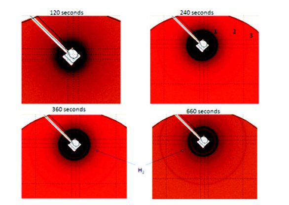
Figure 5 - Synchrotron SAXS scattering from a dispersed soybean oil emulsion over time on exposure to pancreatin.
These experiments show the benefit of using synchrotron SAXS to obtain an understanding of the kinetics and fine detail of the self assembly process in digesting lipid systems that would not otherwise be obtained. The results also indicate that the formation of nanostructure is not predictable, and there is a need to understand determinants and kinetics of nanostructure formation on digestion of lipids in order to inform a future model or framework.
Further Developments
The ultimate purpose of lipid-based drug delivery systems and functional foods is to maintain drug or nutraceutical components in a dissolved state so that they are available for absorption from colloidal structures in the gut. Precipitation of drug during digestion of lipids in vitro has traditionally been an indicator of likely poor performance in vivo. However, many exceptions to this rule have been empirically identified [10], and it is only recently becoming recognized that the solid state properties of the precipitated material are also key to the ultimate utility of these systems; amorphous material is often more readily absorbed than crystalline material due to differences in solubility [11]. Hence establishing the link between colloidal nanostructure and tendency for drug precipitation in vivo would provide an important step forward in the informed engineering of lipid formulations and functional foods. These systems are often further complicated by the presence of surfactants that may or may not themselves be substrates for lipolysis. We plan the application of synchrotron x-ray diffraction, simultaneously with small angle x-ray scattering shown here to link drug solid state properties with colloidal lipid structure in real time – a capability expected to further advance the field of lipid-based formulations for Class 2 compounds. The ultimate use of synchrotron SAXS for in vivo study of the lipid digestion process is also planned.

Figure 6 - Change in nanostructure composition, with changes in initial formulation lipid content
Conclusion
The application of real time synchrotron tyechniques has potential to unlock the links between composition, structure and outcome for lipid-based drug formulations. Rational design of engineered lipid systems to target specific colloidal structures in the gut would enable the field to step beyong empirical formulation design, ultimately leading to improved therapeutic outcomes with BCS Class 2 compounds. (5)
References:
- Hofmann, A., Borgstrom, B. J. Clin. Investig. 1964, 43, 247-257.
- Patton, J., Carey, M. Science 1979, 204, 145-148.
- Porter, C., Trevaskis, N., Charman, W. Nat Rev Drug Discov 2007, 6, 231-248.
- Kossena, G., Charman, W., Boyd, B., Porter, C, J Contr Release 2004, 99, 217-229
- Borne, J., Nylander, T., Khan, A. J. Phys. Chem. B 2002, 106, 10492-10500.
- Borne, J., Nylander, T., Khan, A., Langmuir 2002, 18, 8972-8981
- Fatouros, D., Deen, G., Arleth, L., Bergenstahl, B., Nielsen, F., Pedersen, J., Mullertz, A. Pharm Res 2007, 24, 1844-1853.
- Fatouros, D. G., Walrand, I., Bergenstahl, B., Mullertz, A. Pharm. Res. 2009, 26, 361-374
- Warren, D., Hawley A., Boyd B., Langmuir 2011, 27, 9528–9534.
- Porter, C., Kaukonen, A., Boyd, B., Edwards, G., Charman, W., Pharm Res 2004, 21, 1405- 1412.
- Zhang, F., Aaltonen, J., Tian, F., Saville, D., Rades, T. Euro. J. Pharm. Biopharm. 2009, 71, 64-70.
Author Biography
Associate Professor Ben J. Boyd is a colloid chemist with research interests in colloidal structure in lipid systems and controlling self- assembly to improve drug delivery outcomes. He is Group Coordinator for Drug Delivery Sciences at the Monash Institute of Pharmaceutical Sciences at Monash University in Melbourne, Australia. He has published over 70 journal articles, six book chapters and is President of the Australian Chapter of the Controlled Release Society, and is a member of the Board of Scientific Advisors of CRS.
[email protected]