School of Pharmaceutical Sciences
School of Pharmaceutical Sciences
School of Pharmaceutical Sciences
Introduction
Bioanalysis concerns the analysis of xenobiotics (such as drugs) and endogenous compounds (such as biomarkers) in biological systems. Both xenobiotics and biomarkers encompass low molecular weight (LMW) and high molecular weight (HMW) analytes. According to the analytical purpose, the sample can follow two distinct types of methodologies: untargeted and targeted analysis. The former approach (untargeted) is mainly applied to obtain a maximum of information from the sample to isolate inter- or intra-individual variations and identify potential biomarkers. After validation of a new biomarker, the goal is usually towards quantitation with a maximum of sensitivity, selectivity, and repeatability, as required in targeted analysis. Regardless of the type of analysis, a typical bioanalytical workflow comprises sampling and sample preparation prior to analysis, data treatment, and biological interpretation (Figure 1). Sampling and sample preparation are the first stages of the bioanalytical process. Sampling comprises collection, handling, delivery, processing, and storage of the biological sample prior to sample preparation (Figure 2). Sample preparation is used to clean-up and/or enrich the sample before analysis to improve its detection without fouling the analytical device. According to the nature, availability, volume, and stability of the biological fluid, as well as the goal of the analysis (i.e., targeted or untargeted) and other constraints (e.g. throughput, cost), different sample preparation should be used (Figure 3).
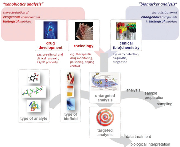 Figure 1. Three main criteria have to be considered to select a sample preparation approach in bioanalysis: (i) the type of analytes (LMW vs. HMW compounds), (ii) the type of biofluid (e.g. blood vs. urine vs. lachrymal fluid), and (iii) the type of analysis (untargeted vs. targeted).
 Figure 2. Pre-analytical parameters can seriously affect bioanalysis.
 Figure 3. Sample preparation techniques according to their respective selectivity and enrichment capabilities. The sphere volumes are proportional to the cost and complexity of the approach.
In untargeted analysis, the main purpose is identification and quantitation of various compounds of interest as example to (i) increase knowledge about a biological pathway, (ii) find new biomarkers, or (iii) determine toxic exposure to an exogenous compound (e.g. illegal drug consumption, toxicant ingestion). Thereby, sample preparation in the context of untargeted analysis needs to be sufficiently versatile and/or generic to avoid information loss, as afforded by non-selective approaches. Extraction procedures can also be used in untargeted analysis because they allow for high sample cleaning, while being not too highly specific towards a single analyte, biological class, or physicochemical property. According to the experimental conditions and the involved interactions, numerous compounds from a common family can be extracted. For extended untargeted analysis, various extraction procedures, exhibiting orthogonal selectivity can be combined to recover a maximum of compounds, together with low contamination from interfering compounds.
In targeted analysis, because the main purpose is the quantitation of a restricted number of analytes or a specific compound, the sample preparation methods used for untargeted analysis may apply but more selective sample preparations, based on highlyspecific recognition, can be advantageous. The choice of the sample preparation is thus very important, since sample identification and quantitation is affected, and therefore results biased, depending on the option selected. All these considerations apply regardless of the field of applications, either in drug development study, toxicological analysis, or clinical (bio)chemistry assays (Figure 1).
Nowadays, there is a growing interest in microextraction (ME) techniques for sample preparation, which may be used in the case of minute amounts of scarce matrices such as lachrymal fluid, cerebrospinal fluid or saliva. Such approaches are also interesting for conventional biofluids such as blood matrices and urine, because they can offer high enrichment when used on large volumes, together in a context of green chemistry due to the low solvent consumption.
This review will focus on alternative sample preparation methods, including ME, which are reported according to the selectivity afforded (Figure 3). Emphasis will also be made on pre-analytic considerations.
Pre-Analytical Aspects
In the past decades, a great effort was made towards improvement in sensitivity, selectivity, and repeatability of bioanalytical methods, and was mainly driven by advances in analytical instrumentation. All these efforts are vain if the sample quality is poor due to pre-analytical errors. Numerous preanalytical parameters may seriously affect the preparation and analysis of biological samples, including factors prior to sample collection (e.g. study design), subsequent collection, handling and delivery to the lab, processing, and inadequate conditions for sample storage (Figure 2).1
The collection and storage of biological samples and associated health information are now well organized in biobanks. These collections of biological samples are particularly relevant in the era of personalized medicine and medical innovations. In biobanks, samples could be associated with clinical data (such as blood pressure or lung volume) and information regarding personal lifestyle (e.g. sport habits or smoking behavior) or can be associated to specific patient groups (e.g. with a specific cancer diagnosis). Therefore, biobanks give access to researchers to samples and data to support medical research, allowing development or confirmatory experiments for new diagnostics, biomarkers, and pharmaceutical treatments. Low volumes of biological matrices can now be collected and readily stored in biobanks under appropriate conditions. Freezing at -80° is recognized as the best temperature compromise in terms of preservation and cost, although -130°C may be needed in some cases.
An alternative for blood collection and storage is the use of dried blood spots (DBS). DBS consists of collecting a few microliters of whole blood drawn by a lancet from the finger, heel or toe and directly blotted on an adsorbent filter paper. The spot is air-dried for a few hours and can be shipped easily and stored at room temperature. Prior to analysis, the dried spot is punched out and the analytes are recovered with an appropriate solvent. Recent automation solutions extract the sample by flushing the elution solvent directly through the filter without punching it out. DBS offers advantages over other sampling methods because it (i) needs minute amounts of biofluid (ca. 10–50 μL), (ii) is less invasive than conventional blood collection, (iii) overcomes difficulties associated with regular sampling, such as special equipment to handle the samples, or fridges and freezers for storing and transport, and (iv) improves the long-term stability of analytes.2 The volumetric bias associated with blood hematocrit is circumvented with recent commercial solutions involving the collection of a controlled volume (regardless of blood viscosity) using micro-channels, which absorb the blood by capillarity and spots accurate volume of blood onto the card. However this format still suffers from its incompatibility with biobanks, where aliquots of real matrices are stored and available for further consideration. A recent alternative uses the same microsampling technology for the rapid generation of plasma from whole blood, giving rise to the so-called dried plasma spot (DPS).3 This technology is now further adopted for other and relevant biofluids, for which only a few microliters are accessible (e.g. saliva, cerebrospinal fluid, lacrymal fluid).4
Sample Preparation for Bioanalysis
Non-selective approaches
For a maximal information content, the most straightforward technique is the dilute & shoot (D&S) approach.5-7 This method consists of diluting the biological sample in an appropriate solvent (according to the subsequent analytical method), to minimize the matrix effects, as well as clogging and fouling of the instrument. For protein content biofluids, a step of protein precipitation (PP) is usually included in the dilution step to alleviate matrix effects from protein content, adsorption to the analytical device, and carry-over. Typical PP are performed by diluting sample in a highly acidic solution (e.g. perchloric acid, trichloroacetic acid), in an organic solvent (e.g. acetonitrile, methanol), or with mineral salts (e.g. zinc sulfate, ammonium sulfate). After PP the filtrate is centrifuged and a small amount of the supernatant is directly injected in the analytical device with or without pH adjustment, or concentrated by solvent evaporation when sensitivity is insufficient. This approach can be applied to a wide variety of compounds. Another feature is the high throughput, which can be further enhanced using 96-well plates format, including proteins’ filtration, as well as phospholipids’ depletion. In some cases, the D&S and PP approaches are not sufficient and despite dilution of interfering compounds, matrix effect can still be very important, particularly when electrospray ionization (ESI) is considered with MS detection. The analytical system can be fouled with the multiple injection of biological samples, giving rise to analytical drift. Another drawback of this approach is the dilution of the analytes themselves, involving that a highly sensitive analytical method, such as LC-, GC-, or CE-MS(/MS) should be used afterwards. With PP, the analyte recovery can be poor due to co-precipitation with the proteins. In this context, sample preparation with higher selectivity and enrichment capability are often recommended.
Solid-based extraction approaches
Solid phase extraction (SPE) is the most commonly used solid-based sample preparation technique in bioanalysis. SPE is based on the analyte interaction between a stationary phase and the sample. A large variety of stationary phases (e.g. reverse phase, normal phase, ion exchange) and numerous formats, from few milligrams to micrograms, are available.
Improvements have been made towards miniaturization and many solid-based ME exist as shown in Figure 4.8 Among them, solid phase microextraction (SPME) consists of a stationary phase grafted on a probe. As for SPE, a large variety of stationary phases and probe dimensions are available. Alternatives to the probe are also used, with syringe needles for instance, as found in microextraction by packed sorbent (MEPS).9 The principal assets of these techniques are (i) application to volatile compounds by head space extraction, (ii) ready hyphenation with GC, and (iii) probe recycling by thermal desorption. Recently, reports have been made using SPME for in vivo sampling: SPME probe was surgically inserted in tissues and organs (e.g. liver, brain) enabling a rapid monitoring of a treated area without tissue lesions.10-11
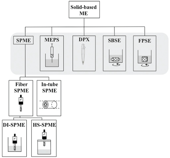 Figure 4. Solid-based microextraction techniques used in bioanalysis. Adapted from,8 with permission.
Other interesting SPE formats have been developed. The first one is the zip-tip format, in which the stationary phase is contained in the end of a 10 μL pipette tip. It has been particularly used in RP mode in the proteomics field for peptide desalting and enrichment. With disposable pipette extraction (DPX), pipette tips incorporate loosely contained sorbent material, which is mixed with the sample solution. Turbulent air bubble mixing creates a suspension of sorbent in the sample ensuring optimal contact and efficient extraction.
In order to improve the mass transfer and the extraction speed, stir bar sorptive extraction (SBSE) and fabric phase sorptive extraction (FPSE) have been developed. In SBSE, the stationary phase is grafted on the surface of a magnetic bar, which is deepened in the sample, whereas in FPSE the stationary phase is bonded to a permeable fabric (e.g. cotton, paper). The main advantage of these approaches is the improvement of the contact surface between the sample and the stationary phase, leading to high recovery, high enrichment, in a minimal time, and fewer extraction steps.
In the proteomics field, the HMW compounds of interest can be at a very low abundance compared to high abundant proteins. Extraction of the former can hardly be achieved by conventional SPE procedure due to their close behavior compared to the latter. Depletion approaches have thus been developed to allow for minor HMW compounds expression. They are based on selective interactions between the proteins and random hexapeptides grafted on beads used as ligand: theoretically each hexapeptide bind to a unique protein. Because of the bead capacity loading limits, major proteins will quickly saturate their ligand and the excess will be washed. On the other hand, minor proteins will be concentrated on beads prior to elution of all the retained proteins.12 Based on a similar mechanism, (i.e. antibody recognition) immunodepletion, allows suppressing major contaminant proteins from a sample. Immunodepletion columns are commercially available in microcartridge format and can deplete up to 22 major proteins from the plasma.13
Liquid-based extraction approaches
Liquid-liquid extractions (LLE) are interesting alternatives to solidbased approaches. In LLE, extraction occurs according to the lipophilicity of the compound of interest and the partition takes place between an immiscible organic solvent and the biological sample. Conventional LLE uses large amounts and sometimes toxic organic solvents. In order to enhance recovery, enrich the sample, and reduce extraction time and organic solvent consumption, different improvements have been developed with ME. As reported in Figure 5, liquid-based ME are derived either from single-drop microextraction (SDME), in which a single drop of water-immiscible solvent suspended from the tip of a syringe is immersed in the aqueous sample, or hollow fiber liquid-phase microextraction (HFLPME), in which a hollow polymeric fiber is used as a support for the acceptor phase.
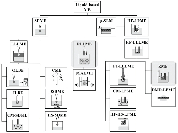 Figure 5. Liquid-based microextraction techniques used in bioanalysis. Adapted from,8 with permission.
Dispersive liquid-liquid microextraction (DLLME) belongs to the first family and enables high surface contact between the two immiscible phases by creating a microemulsion using a dispersive solvent.14 A quick equilibrium state is achieved, followed by a centrifugation step to break the microemulsion. Because the recovered solvent is often nonmiscible with water and therefore incompatible for a RP-LC injection, evaporation is usually performed and the extracted compounds are recovered in a low volume of compatible solvent, allowing for high enrichment.15-17
Another remarkable liquid-based ME, from the second family, is electromembrane extraction (EME).18 A typical EME device is composed by two compartments, the donor compartment, which is the biological sample, and the acceptor compartment. They are separated by a polymeric membrane impregnated by an organic solvent immiscible with water, also called supported-liquid membrane (SLM). An electric field is applied between the two compartments, enabling migration of compounds through the SLM. With this approach, passive diffusion is not the only phenomenon but an active migration occurs and high recovery can be obtained in a very short time. Two types of selectivity is afforded by EME: (i) according to the electric field polarity, anions or cations are preferentially extracted and (ii) according to the SLM composition (i.e., nature of the organic solvent and presence of carrier compounds), analytes with different lipophilicity are preferentially extracted. While EME has been widely used for basic and acidic LMW drugs, it still remains a challenge for HMW analytes such as peptides, due to their low mobility and lipophilicity.19
Other approaches have been developed for polar compounds, to reach higher recoveries than that obtained in conventional LLE. Saltassisted liquid-liquid extraction (SALLE), already used in QuEChERS protocols for pesticide analysis,20,21 uses polar organic solvents such as acetonitrile or alcohols. The principle is based on the salting out effect, which in high salt concentration, renders water immiscible with ordinarily water miscible organic solvents. SALLE can be coupled with PP, allowing for the extraction and the elimination of interfering proteins in a single step.
Highly-selective approaches
Immunoprecipitation (IP) is based on the recognition between a specific monoclonal or polyclonal antibody (Ab) and its ligand. Usually, Ab are immobilized on a solid support such as a microcartridge or magnetic beads. With this approach, very high selectivity and high enrichment are obtained, particularly for HMW compounds such as proteins. It should be noted that Ab selection, optimization, and production are time-consuming and costly. A similar approach based on oligosorbent uses the recognition between a ligand and immobilized aptamers, rather than Ab. Aptamers are made of short single-stranded oligonucleotides (20 to 100 unities) presenting a unique three-dimensional structure able to bind a large variety of ligands.22 As for IP, the development can be tedious due to the different steps for aptamer selection and enrichment by polymerase chain reaction (PCR).23-24 Unlike IP though, aptamer production does not require any living organisms, is cheaper, and more repeatable. Both techniques feature high selectivity towards HMW compounds due to their 3D structure and high number of recognition sites. For LMW analytes, lower selectivity is observed due to the close structures between small chemical compounds.
Another approach is based on molecular imprinted polymers (MIP). This technology uses a molecule mold (size, shape, surface chemistry) in a polymeric matrix, which is obtained by mixing the targeted analyte or a structural mimetic compound in an appropriate mixture composed of a monomer. During the polymerization reaction, the polymer freezes the orientation of the functional groups. After grinding of the polymeric matrix, fragments can be packed in a microcartridge or included in a pre-column. Recognition between the polymeric mold and the targeted analyte is made by non-covalent interactions (e.g. electrostatic, dipole-dipole, van der Walls).25 Although this technology is cheaper and easier to develop than IP methods, the optimization of MIP and its synthesis can be long and requires large amounts of targeted analyte or mimetic. While real applications in biological sample preparation are still awaited, other application fields are starting to make use of MIP, in drug discovery and drug delivery applications for instance.
Conclusion
Sampling and sample preparation are the first steps encountered in bioanalysis and are crucial to ensure appropriate biological interpretation. Because scarce biological fluids are now of growing interest, microextraction techniques have been developed to enable handling of low volumes of samples and work in agreement with the concept of green analytical chemistry.
References
- Yin, P., R. Lehmann, and G. Xu, Effects of pre-analytical processes on blood samples used in metabolomics studies. Anal Bioanal Chem, 2015. 407(17): p. 4879-92.
- Deglon, J., L.A. Leuthold, and A. Thomas, Potential missing steps for a wide use of dried matrix spots in biomedical analysis. Bioanalysis, 2015.
- Li, W., et al., Evaluation of plasma microsampling for dried plasma spots (DPS) in quantitative LC-MS/MS bioanalysis using ritonavir as a model compound. J Chromatogr B Analyt Technol Biomed Life Sci, 2015. 991: p. 46-52.
- Sen, A., et al., Metabolic phenotype of the healthy rodent model using in-vial extraction of dried serum, urine, and cerebrospinal fluid spots. Anal Chem, 2013. 85(15): p. 7257-63.
- Dong, Y., et al., A Sensitive Dilute-and-Shoot Approach for the Simultaneous Screening of 71 Stimulants and 7 Metabolites in Human Urine by LC-MS-MS with Dynamic MRM. J Chromatogr Sci, 2015.
- Cao, Z., E. Kaleta, and P. Wang, Simultaneous Quantitation of 78 Drugs and Metabolites in Urine with a Dilute-And-Shoot LC-MS-MS Assay. J Anal Toxicol, 2015. 39(5): p. 335-46.
- Rodin, I., et al., ‘Dilute-and-shoot’ RSLC-MS-MS method for fast detection of nerve and vesicant chemical warfare agent metabolites in urine. J Anal Toxicol, 2015. 39(1): p. 69-74.
- Kohler, I., J. Schappler, and S. Rudaz, Microextraction techniques combined with capillary electrophoresis in bioanalysis. Anal Bioanal Chem, 2013. 405(1): p. 125-41.
- Silva, C., et al., Microextraction by Packed Sorbent (MEPS) and Solid-Phase Microextraction (SPME) as Sample Preparation Procedures for the Metabolomic Profiling of Urine. Metabolites, 2014. 4(1): p. 71-97.
- Bojko, B., et al., Low invasive in vivo tissue sampling for monitoring biomarkers and drugs during surgery. Lab Invest, 2014. 94(5): p. 586-94.
- Cudjoe, E., et al., Solid-Phase Microextraction: A Complementary In Vivo Sampling Method to Microdialysis. Angewandte Chemie International Edition, 2013. 52(46): p. 12124-12126.
- Thulasiraman, V., et al., Reduction of the concentration difference of proteins in biological liquids using a library of combinatorial ligands. ELECTROPHORESIS, 2005. 26(18): p. 3561- 3571.
- Staub, A., et al., Analysis of hemoglobin-based oxygen carriers by CE-UV/Vis and CE-ESITOF/ MS. Electrophoresis, 2010. 31(7): p. 1241-7.
- Rezaee, M., et al., Determination of organic compounds in water using dispersive liquid– liquid microextraction. Journal of Chromatography A, 2006. 1116(1–2): p. 1-9.
- Zuloaga, O., et al., Dispersive liquid-liquid microextraction: trends in the analysis of biological samples. Bioanalysis, 2015. 7(17): p. 2211-25.
- Li, Y., et al., Development of a Efficient and Sensitive Dispersive Liquid-Liquid Microextraction Technique for Extraction and Preconcentration of 10 beta2-Agonists in Animal Urine. PLoS One, 2015. 10(9): p. e0137194.
- Kohler, I., et al., Dispersive liquid-liquid microextraction combined with capillary electrophoresis and time-of-flight mass spectrometry for urine analysis. J Pharm Biomed Anal, 2013. 73: p. 82-9.
- Gjelstad, A., S. Pedersen-Bjergaard, and K.F. Seip, Electromembrane extraction as a rapid and selective miniaturized sample preparation technique for biological fluids. Bioanalysis, 2015. 7(17): p. 2203-9.
- Huang, C., A. Gjelstad, and S. Pedersen-Bjergaard, Exhaustive extraction of peptides by electromembrane extraction. Anal Chim Acta, 2015. 853: p. 328-34.
- Valente, I.M. and J.A. Rodrigues, Recent advances in salt-assisted LLE for analyzing biological samples. Bioanalysis, 2015. 7(17): p. 2187-93.
- Xiong, X. and L. Yang, Salting-out-assisted liquid-liquid extraction with acetonitrile for the determination of trimetazidine in rat plasma using liquid chromatography-mass spectrometry. Biomed Chromatogr, 2015. 29(2): p. 268-74.
- Stoltenburg, R., C. Reinemann, and B. Strehlitz, SELEX—A (r)evolutionary method to generate high-affinity nucleic acid ligands. Biomolecular Engineering, 2007. 24(4): p. 381-403.
- Mascini, M., I. Palchetti, and S. Tombelli, Nucleic acid and peptide aptamers: fundamentals and bioanalytical aspects. Angew Chem Int Ed Engl, 2012. 51(6): p. 1316-32.
- Pichon, V., F. Brothier, and A. Combes, Aptamer-based-sorbents for sample treatment--a review. Anal Bioanal Chem, 2015. 407(3): p. 681-98.
- Schirhagl, R., Bioapplications for molecularly imprinted polymers. Anal Chem, 2014. 86(1): p. 250-61.
Biography
Nicolas Drouin is pharmacist from the University of Lorraine and master graduate in Analytical Sciences from the University of Strasbourg with a master thesis in mass spectrometry proteomics. Since 2013, he is PhD student at the University of Geneva. His studies involve the development of new sample preparation methods based on electromigration for the analysis of scarce biofluids.
Serge Rudaz is associate professor at the University of Geneva. He is research group leader at the Swiss Centre for Applied Human Toxicology and member of various scientific boards. He published numerous scientific papers (>230) and is currently interested in toxicological analysis, including counterfeit medicines, bioanalysis, and metabolomics.
Julie Schappler is pharmacist and holds a PhD from the University of Geneva. She is currently senior lecturer and research associate. She heads the unit of capillary electrophoresis and sample preparation, which works on developing methods to improve analysis performance while reducing analysis time and cost. Applications range from small pharmaceutical compounds and their metabolites in biofluids, to biomolecules.