More than 100 scientists met in Ulm at the end of September for the 14th Confocal Raman Imaging Symposium. The lectures thoroughly documented how modern Raman microscopy contributes to gaining new insights into widely varied scientific questions.
In 11 invited lectures three short presentations, the speakers presented new and interesting applications of Raman microscopy to answer questions from life sciences, materials science, geoscience and environmental science.
After an overview of the physics of the Raman Effect with Sebastian Schlücker (University of Duisburg-Essen, DE), Admir Masic from MIT (Cambridge, USA) reviewed his studies of biological and archaeological materials, such as the teeth of sea urchins, Roman concrete or 2000-year-old animal hide, which was used as writing material at that time. Masic said he wanted to use his knowledge of structures and design principles to develop new materials and processes.
In the life sciences session, Maike Windbergs (Goethe University, Frankfurt, DE) described how Raman microscopy can be used to investigate cellular in vitro models that are used in pharmaceutical research as a substitute for animal experiments. For example, she wants to find out whether the active ingredients of generic drugs behave in exactly the same way as those of the original compounds and what influence formulations have on the effect of the substances. Paul Pantano (University of Texas, Dallas, USA) also presented studies on the inner life of cells. He monitors whether and how nanoparticles are absorbed by them. As they’re a particularly Raman-active test material, he uses carbon nanotubes (CNTs). Five lectures were part of the Materials Science session. Satender Kataria (RWTH Aachen University, DE) and Siegfried Eigler (Free University of Berlin, DE) presented their Raman studies on 2D materials. Kataria described graphene-based pressure sensors and photodetectors he designed. Eigler deals with the functionalization of graphene and described how such processes can be tracked with the help of Raman spectra. Bastian Barton (Fraunhofer Institute for Structural Durability and System Reliability, Darmstadt, DE) is working on automating Raman microscopy for the analysis of multilayer polymer films.
Helena Nogueira (University of Aveiro, PT) is involved in the analysis of textile fibers. She uses Raman microscopy to investigate the distribution of silver particles and dyes on fibers used in the production of functional textiles. José Swart (AkzoNobel Chemicals, Deventer, NL) presented very practical applications. Not only has she put wall paints and anti-rust coatings under the Raman microscope, but she’s also examined how quickly antifouling substances are washed out of the underwater coatings of boats.
Raman microscopy is also an important method for geoscientists and environmental scientists. Natalia Ivleva (Technical University of Munich, DE) uses it for the analysis of microplastics in water. These particles are considered harmful to the environment. Dina Bower (NASA Goddard Space Flight Center, Greenbelt, USA) identifies Raman signatures of organic life in rocks billions of years old, to later help in the search for such signatures far beyond Earth.
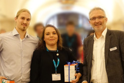
Winners of the Poser Award Thomas Rabl (left) and Vera Duganžic with WITec’s CEO Joachim Koenen (right).
The three short presentations also reflected the diversity of the world of Raman microscopy. Jens Sommertune (RISE Bioscience and Materials, Stockholm, SE) provided insight into his life as a "Raman Service Scientist." He’s already had samples as diverse as rust, cheese and corena under the microscope. Zuzana Kroneková (Polymer Institute of the Slovak Academy of Sciences, Bratislava, SK) is investigating how microcapsule systems release pharmaceutical agents. Her aim is to pack insulin-producing cells in tiny polymer capsules and implant them into diabetic patients.
Walter Müller (Friedrich Schiller University, Jena, DE) posed a question that was exciting for the entire Raman community: Can Raman microscopy be accelerated? That would be of particular interest for the analysis of living cells, but also for high-throughput investigations. If one wants to measure faster, an increase in laser power is necessary, which is difficult for living cells and organisms to withstand. Müller compared Raman microscopes with different types of illumination and calculated the thermal load on the samples. In lightsheet microscopy, in contrast to confocal microscopy, illumination can capture the entire spectral information of the field of view at the same time, but this illumination geometry leads to heat build-up in the sample, which can only be avoided by a significant reduction in the excitation intensity.
As in previous years, the best posters were recognized with awards in 2017. The jury chose to award two works, both from the field of medicine/pharmaceutical research. Chemist Vera Duganžic (Leibniz Institute for Photonic Technologies, Jena, DE) showed studies for the detection of macrophages in atherosclerotic plaques using modified gold particles. The presence of these immune cells is considered as indication of an approaching dangerous rupture of the plaque that triggers thrombosis. Thomas Rabl (University of Dundee, UK) showed how he can determine the concentration of antiparasitic drugs in human cells with the help of silver particles. The better the cellular properties of such a therapeutic agent, the stronger it can accumulate in immune cells and the more effective the defense against parasites can be. This method of investigation could prove helpful in the development of new antiparasitic drugs.
At the end of the symposium, the participants were invited to WITec Headquarters in Ulm, where they were shown the latest confocal and correlative Raman microscopes and developments in software. A survey at the end of the event revealed that the participants felt very well informed and were inspired by the diverse applications of this type of spectral microscopy.