Introduction
An important area for the advancement of pharmaceuticals is in product formulation development. The active pharmaceutical ingredient (API) delivery method can be as important as the drug itself. Hot melt extrusion (HME) is a method that has been used quite extensively in the plastics industry to produce a wide assortment of plastic products in a variety of forms. HME applied to the production of pharmaceutical formulations is a relatively new method being developed as an attractive alternative to traditional processing methods for producing products such as tablets, granules, pellets, and even transdermal films.
The HME process has 3 primary advantages over traditional methods. First, HME is an efficient manufacturing method that can be run as a continuous process, allowing for direct in-line monitoring and control over the manufacturing process. Secondly, HME is an easier way to produce a variety of different dosage formulations. Finally, HME offers a means to create products with enhanced dissolution, improved product delivery, and controlled release.
The lack of solvents in HME processing saves money and eliminates steps such as drying. The dispersion of APIs in thermoplastics can lead to different therapeutic activity. HME can produce thermodynamically stable solid solutions and suspensions that extend product lifetimes.
In a simplistic view of the HME manufacturing process, the API and other components are combined with pharmaceutically approved thermoplastic polymers at elevated temperatures (50°C to 180°C). Rotating screws are typically used for mixing the components and conveying them to the extrusion die. The screw threads are specifically designed to control the mixing and transport properties of the materials at the various stages in the process. The temperature is controlled for uniform passage of the molten material through the extrusion die. The final form depends on the die and any post extruder processing.
Advanced feeders for raw materials, exacting control over the mixing (shear) and heating processes, and monitoring of the extruded product are all essential for producing the desired product. It is important to recognize how the different processing conditions affect the API and excipients. Verifying the distribution and integrity of the various components helps assure a reliable and reproducible product.1
Analytical Methods for Evaluating HME Products
Fast and easy-to-use analytical methods are necessary to understand the formulation process, the effect of the processing conditions, and to monitor the quality of the product. While near infrared (NIR) spectroscopy is an excellent choice for process monitoring,1 Raman micro-spectroscopy is a better choice for detailed evaluation of component distributions. Raman spectroscopy can be used to identify and verify the presence of different components and contaminants in various formulations. However, the utility of Raman spectroscopy is not limited to just identification of materials; it also provides detailed information on molecular structure and chemical environment which reveal subtle differences in the structure and orientation of molecules. Polymorphs and solvates can be differentiated as well as the physical properties of materials such as stress or degree of crystallinity.
Single point Raman analysis is a powerful tool for examining materials but Raman imaging opens up a new way of looking at samples. Imaging provides views of the spatial distributions of components and the variation of physical properties throughout the sample. The vast amounts of spectroscopic data produced using Raman imaging are used to create images highlighting different chemical and structural aspects of the components in the sample. These images allow for a quick visual analysis of the samples.
Experimental Details and Samples
A Thermo Scientific™ DXR™xi Raman imaging microscope and Thermo Scientific™ OMNIC™xi software were used for acquiring and processing the Raman imaging data presented in this paper. OMNICxi software allows for easy optimization of collection parameters and provides options for choosing single, multiple, or even auto-selected regions. The DXRxi Raman imaging microscope collects Raman spectral data at extremely fast rates. This means that the collection of large area Raman images is now not only practical, but routine. The DXRxi Raman imaging microscope still retains the best qualities of the original DXR Raman microscope but has a new state-of-the-art, high speed microscope stage-synchronized with a highly sensitive EM CCD detector, which provides this instrument with the power to accurately and reliably collect a large amount of data in a small amount of time. The OMNICxi software provides a straightforward graphic interface for defining regions and collecting data, and provides the analysis tools to generate meaningful Raman images based on a variety of spectroscopic profiles.
The HME samples detailed in this paper were provided by Dr. Adrian Kelly from Bradford University, UK. They were created using a Thermo Scientific™ Pharmalab 16 twin screw hot melt extruder. Hypromellose acetate succinate (HPMCAS) was used as the soluble polymer carrier, and the API was ibuprofen (25% to 33%). Some formulations also contained D-mannitol (7% to 15%). They were supplied as cross-sections of extruded material mounted in epoxy resin. The goal was to evaluate the spatial distribution of the components and to ascertain if the processing conditions caused any unforeseen changes.
Ibuprofen is typically present in pharmaceutical formulations as a racemic mixture of 2 stereoisomers. Besides the 2 different enantiomers, there have also been reports of ibuprofen existing in other polymorphic forms.2 Phase I is thermodynamically stable while phase II is an uncommon, meta-stable form that has been generated by rapid quenching of molten ibuprofen. The Raman spectrum of amorphous glassy ibuprofen is subtly different from the crystalline form.3 There have also been reports of ibuprofen co-crystallizing with other materials such as nicotinamide during hot melt extrusion.4 Changes in the Raman spectra of ibuprofen when forming extrudates with other carriers such as polyvinyl pyrrolidone (PVP) have also been observed.5 The polymorphic phases and the molecular associations are examples of materials that could be differentiated using Raman imaging. While not all of these differences will noticeably affect the utility of the pharmaceutical formulation, it is still good to understand the changes that may or may not be occurring during the hot melt extrusion process.
Raman Imaging Results
The Raman image of the entire cross-section of one of the extruder samples is shown in Figure 1. In this case, the area analyzed was approximately 6.35 mm x 4.6 mm and a 10x objective was used to collect both the visual image and the Raman data. This area includes some of the epoxy around the sample. The image pixel size in this case is 25 microns giving an image based on 46736 spectra. A 780-nm laser (24 mW) was used to avoid the significant fluorescence observed with shorter wavelength lasers.
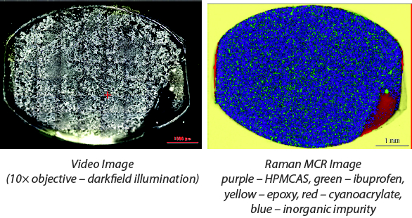 Figure 1. 10× objective, 780-nm laser, 24 mW, 6.3 × 4.6 mm area, 25 mm image pixel size, 46736 spectra, 0.0100 s exposure time, 100 scans.
The image is the result of a multivariate curve resolution (MCR) analysis of the spectroscopic data. MCR differentiates the Raman spectra into components; the constituents are assigned different colors to produce a chemical image and identified using automatic library searching routines. In this case, the HPMCAS is purple, ibuprofen is green, and the mounting epoxy is light yellow. An image analysis of the green component (ibuprofen) indicates that it is 23% of the HME product area. This is close to the percentage of ibuprofen reported in the original formulation at 25%. There is some variability in this type of analysis and, while there are some limiting assumptions, this area analysis provides a rough idea of the relative amounts of the components in the sample. Spectra from the area shown in red displayed a prominent nitrile peak, and the spectra are consistent with a cyanoacrylate impurity. The blue color indicates an area where the material is mostly HPMCAS, but has 2 additional broad peaks present (606 and 454 cm-1) which may indicate an inorganic impurity, possibly some type of titanate. These results demonstrate the ability of the MCR routine to very clearly differentiate and highlight unexpected components even when present in relatively low amounts.
A closer look at the sample reveals interesting detail not readily apparent at the lower magnification. Figure 2 includes both visual and Raman images obtained using a 50x objective. The Raman image was collected at higher resolution (3 μm image pixel size). A smaller representative portion of the sample (687 mm x 423 mm) was analyzed but as a result of the higher resolution this image still contained over 32000 spectra. While it is possible to image the entire sample at higher resolution, it isn’t necessary. The more effective and efficient option would be to use the multiple region option in the OMNICxi software to target the most interesting portions of the sample.
 Figure 2. 50× objective, 780-nm laser, 24 mW, 687 × 423 mm area, 3.0 mm image pixel size, 32289 spectra, 0.0100 s exposure time, 100 scans.
The higher resolution image shows significantly greater detail and the MCR analysis identifies 2 different components as ibuprofen. A review of the spectra from these 2 areas (green and yellow) showed significant differences in the ibuprofen spectra (see Figure 3). These manifest themselves as variances in relative peak intensities and shapes. The most pronounced differences are highlighted in Figure 3 and are consistent not only in other images collected from this sample but other HME samples as well. The spectral variations are inconsistent with those reported for the polymorphs of ibuprofen, glassy ibuprofen, or for co-crystalization products. While the cause of these spectral variations has not been established at this time, a possible explanation might involve orientation differences of small crystallites of ibuprofen but that needs to be investigated further.
 Figure 3. Spectral differences: representative Raman spectra of ibuprofen from the green and yellow areas of the MCR image compared to a library spectrum.
Figure 4 shows a Raman image of one of the HME products containing mannitol and ibuprofen. Although the sample image is similar to those previously observed, in this case mannitol was identified as an additional component. The spatial distribution revealed some sizable particles of mannitol, and it was not uniformly dispersed. The figure insert shows the effect of increased spatial and image resolution. In this case, the increased resolution produced a more visually appealing image but did not provide any additional substantial information, emphasizing the point that higher resolution, while at times very useful, is not always required. It is often more efficient to reserve highresolution analysis for specific targeted regions of the sample.
 Figure 4. Raman MCR image: blue – HPMCAS, green – ibuprofen, orange – mannitol, yellow – epoxy, fuchsia – cyanoacrylate.
Conclusions
HME is an innovative way of formulating pharmaceutical products offering a variety of advantages over traditional methods including more control over dosages and forms of the products. However, new technology creates new challenges as well. It is necessary to understand how these new processing methods affect components, and to verify the distribution of the components within the HME products. Raman imaging not only provides a way to identify and verify components but it also provides visual representations of spatial distributions. This visual analysis can be extended to include images based on differences is state, such as solvation, degree of crystallinity, polymorphism, molecular association, and co-crystalization. The DXRxi Raman imaging microscope and OMNICxi imaging software are a powerful combination that provide high-quality imaging results with an-easy-to use, straightforward user interface making Raman imaging assessable for any application.
References
- Chirkot T, Halsey S.Swanborough A. Monitoring the Output of Pharmaceutical Hot Melt Extruders with Near-infrared Spectroscopy. Application Note 51836, Thermo Fisher Scientific.
- Dudognon E, Correia NT, Daneda F, Descamps M. Solid-Solid Transformation in Racemic Ibuprofen. Pharm Res. 2013;30:81-89.
- Hedoux A, Guinet Y, Derollez P, Dudognon E, Correia NT. Raman spectroscopy of racemic ibuprofen: Evidence of molecular disorder in phase II. International Journal of Pharmaceutics. 2011;421:45-52.
- Dhumal RS, Kelly AL, York P, Coates PD, Paradkar A. Cocrystalization and Simultaneous Agglomeration Using Hot Melt Extrusion. Pharm Res. 2010;27:2725-2733.
- Breitenbach J, Schrof W, Neumann J. Confocal Raman-Spectroscopy: Analtsical Approach to Solid Dispersions and Mapping of Drugs. Pharmaceutical Research. 1999;16(7):1109-1113.
Author Biography
Dr. Robert Heintz is a Senior Applications Specialist for Thermo Fisher Scientific. He received a BS in chemistry from Plattsburgh State University College in 1987. He earned a PhD from Cornell University in inorganic chemistry in 1994. Dr. Heintz did his post-doctoral work with Dr. Kim Dunbar at Michigan State University. Dr. Heintz has numerous published research papers (25+) including 2 book chapters and a patent.