Introduction
During manufacturing of biopharmaceuticals prokaryotic and eukaryotic protein expression host systems like Escherichia coli (E. coli) and Chinese Hamster Ovary (CHO) cells generate a heterogeneous mixture of the desired therapeutic protein and a variety of endogenous host cell proteins (HCPs) with different physicochemical and immunological properties. HCPs, as the predominant class of process related impurities are undesirable in the purified protein product as they are suspected to have an impact on patients safety.1,2 In extreme cases residual HCPs have the potential to elicit adverse events such as immune response against the individual HCP itself or the formation of anti-drug antibodies.3,4 Under some circumstances HCPs may even be biologically active in the human body or impact product stability by degradation either of the therapeutic protein or product-stabilizing excipients.5,6 Consequently, quantitation and removal of HCPs in the final drug substance (DS) to acceptable low levels is essential. Based on risk assessments, clinical experience and manufacturing capability typically accepted levels are < 100 ppm (100 ng HCP/mg product).4,7-9 To detect such small total quantities of HCPs against the background of protein product being present at huge excess a highly sensitive and specific analytical method with a wide dynamic range is required. Multi-analyte enzyme-linked immunosorbent assays (ELISA) capable of detecting the majority of protein impurities are routinely used to quantify and monitor HCPs. HCP ELISAs are primarily applied as an in-process control to assess product purity at release testing level of a biopharmaceutical and to demonstrate process consistency but are also used to support process development and process validation.10 Depending on the stage of clinical development, different types of HCP immuno-assays are required. While during earlier clinical phases a generic/platform assay may be adequate, health authorities do expect that a product/process-specific assay is employed at late stage development prior to BLA license application, which is backed-up with sufficient data obtained by orthogonal methods. Here, we provide an overview of HCP characterization strategies including qualification of the critical reagents to demonstrate HCP ELISA suitability, the importance of orthogonal analytical methods to demonstrate product purity as well as further requirements (Figure 1). In addition, the advantages and limitations of commonly applied methods will be discussed.
 Figure 1. Characterization strategy for host cell proteins
HCP Assay Qualification – Demonstrating HCP ELISA Suitability
It must be noted that assessments done for HCP assay qualification and demonstration of suitability of HCP assays should be performed based on recently published regulatory requirements.11,12 The performance of immuno-based assays for quantification and monitoring of HCPs in biopharmaceuticals is tightly linked to the quality of the critical reagents. For a multi-analyte HCP ELISA these critical reagents are typically the HCP antigen and the corresponding polyclonal antibodies (antisera).
HCP Reference Standard
The HCP antigen - typically also used as the Reference Standard - for later analytical ELISA testing is produced from a non-transfected or mock-transfected (without product coding gene) cell culture fermentation, using representative bio-process manufacturing conditions. In order to demonstrate that the HCP protein composition of the null cell culture fermentation is representative for the manufacturing process under investigation, a comparative protein analysis by two dimensional difference gel electrophoresis (2D-DIGE) is applied.12 This approach enables simultaneous separation of HCPs that are covalently pre-labeled with different fluorescent dyes in a single gel and yields at least the detection sensitivity of most widely applied silver staining procedures.13-15 By this means, thousands of varying proteins can be routinely separated and compared. The HCP profile comparison of the null cell culture fermentation (reference antigen) and the investigated biologics manufacturing process is typically conducted by manual (visual) or automated (software based) protein spot detection and evaluation. However certain proteins, especially hydrophobic, excessively large or small and extremely acidic or basic proteins can potentially be under-represented or in worse cases not detected at all.14,16 Furthermore, bio-processed proteins may exhibit diverse posttranslational and/or chemical modifications with different biochemical properties resulting in scattered spot-locations on the gel, impeding sample comparison.
A complementary method to characterize the HCP reference standard is liquid chromatography tandem mass spectrometry (LC-MS/MS). After enzymatic digestion of proteins (usually with trypsin), the obtained peptides are separated via high-performance liquid chromatography (HPLC) followed by tandem mass spectrometry (MS/MS) analysis and subsequent protein identification. This powerful technique enables identification and comparison of thousands of individual HCPs between samples. One of the most critical parts in this workflow is mass spectrometric data analysis supported by a specific data-base search. Proteins are identified by a common database search based on the comparison of the experimental raw data spectra and theoretical in silico generated MS/MS spectra. Besides the content/quality of the used database (e.g. partial sequences or redundant entries) it is important to perform data processing with well defined, consistent and documented parameters.17,18
HCP Specific Antibodies
Different isolation strategies can be applied to purify the polyclonal antibodies against HCPs from the raw sera that have been derived from immunized host species (i.e. sheep, goat, rabbit).19 The two major strategies involve affinity purification that can be either achieved with immobilized protein G/A or with immobilized HCPs.20 Regardless of the purification strategy used, it is essential that the polyclonal antibodies recognize a broad range of HCPs including proteins with extreme net charges or molecular sizes. A widespread methodology for determining the quality of the polyclonal antibodies is 2D gel electrophoresis followed by Western Blotting. The most common and established detection technique for bound antibodies is enhanced chemiluminescence (ECL), however more recent approaches involve the application of fluorescence labeled conjugates. Fluorescence detection offers several advantages over chemiluminescence methods including a more stable signal, high linear dynamic range for quantification and the possibility to detect more than one target by using different dyes at the same time. An important read-out of the above-mentioned approaches is the so called coverage, which refers to the ratio of HCPs recognized by the antibody relative to the total HCP population. Irrespective of whether visual or software based coverage determination is applied, it remains a challenge to match total protein stain and immunostaining, as size and shape of spots sometimes show a poor correlation between protein spots and immunogenic spots. Furthermore, spots can be comprised of more than one protein, and individual proteins can be distributed over several spots as a result of post-translational modifications or proteolytic cleavage. Moreover, due to the protein denaturing conditions during 2D SDS PAGE separation, there is an inherent tendency of linear epitopes being preferentially detected out of the HCP antigen.20 In particular, these limitations complicate especially quantitative coverage assessment and calculated numbers for detection coverage should only be regarded as good approximation.
Another important approach for determining detection coverage is affinity based mass spectrometry (native coverage). At first, immobilized polyclonal antibodies are used to capture host cell proteins. Secondly, bound proteins (covered HCPs) are subjected to LC-MS/MS measurements for identification.21,22 This approach not only enables assessing ELISA reagent coverage under native conditions, but also provides a better understanding of the population of HCPs not sufficiently tracked, which in turn facilitates improvement of the ELISA reagents. Still challenges remain with this technology. For example, the short on-column contact time of the antigen-antibody complex may lead to an underestimation of an ELISAs capability to detect weakly bound HCPs and overall robustness of the whole approach needs to be improved.
2D gel electrophoresis followed by Western Blotting is still a valuable and widely accepted tool for coverage determination. However, MS-based detection of affinity purified HCPs may become more important in the future as this technology further improves, mainly due to its native HCP separation approach being more relevant to ELISA assay conditions.
Product Purity Determination – Orthogonal Methods
Targeted quantitation of individual HCP species contributing to the overall signal is not possible by HCP ELISA. The quantitative values obtained by the HCP ELISA represent a cumulative parameter of all the immune-reactive HCPs present in a particular sample in relation to the HCP reference standard. Furthermore, polyclonal anti-HCP antibodies are present in different quantities and show diverse affinity and avidity, limiting the capability of the HCP ELISA to quantify all HCPs with the same sensitivity. This could potentially lead to an over- or underestimation of particular HCPs.20 As a consequence of an extreme underrepresentation or complete absence of specific antibodies against certain HCPs, the latter may not even be detected at all. To overcome this limitation, it is highly recommended to use orthogonal methods. Mass spectrometry provides a universal approach to identify and characterize HCPs without the need for critical reagents like HCP reference standard or antisera.23,24 The liquid chromatography tandem mass spectrometry (LC-MS/MS) technique is capable of addressing the complexity and dynamic range of HCP pools in bio-process samples, typically with a sensitivity in the lower ppm range, enabling detection of high to moderate abundant HCPs in purified drug substance material. To increase the confidence in protein identification achieved by automated database searches, however, the mass spectrometric results generated should be manually reviewed.17,25 In highly purified samples negative findings (no detection of HCPs) are common, and as such a system suitability test (e.g. spiked individual HCP ideally at or close to the detection limit) should be used to verify the system performance.23 Nevertheless, mass spectrometry requires expensive equipment with high mass accuracy and resolution as well as a highly experienced operator and exhibits limited throughput compared to ELISA.20,26
Additional Requirements – Quantification of Individual HCPs
In some cases, single product related lead HCPs may be found to be insufficiently recognized by the standard ELISA. To support a targeted risk assessment for this species, it is highly beneficial to establish a specific assay to enable a sensitive and accurate quantification (Table 1).22 As previously outlined, quantification can be realized either by mass spectrometry based methods or immunoassays. Once established, protein specific immunoassays are highly sensitive, accurate and precise and can be used in a high throughput manner. Nevertheless, the long lead time for implementing such an assay prohibits the application for first troubleshooting activities or to support early bioprocess improvement.
Table 1. For host cell proteins (HCP-X) risk assessment quantification of individual HCP-X identified by mass spectrometry is necessary. Quantification of HCP-X can be realized either by mass spectrometry based methods or immunoassays.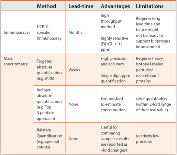
In contrast, mass spectrometry is able to quantify proteins without the need of specific antibodies.27-29 One major approach is targeted tandem mass spectrometry (MS/MS) such as multiple reaction monitoring (MRM) or parallel reaction monitoring (PRM) methods. In these techniques a predefined list of proteolytic peptides and fragment ions are monitored. Recombinant protein or stable isotope labeled peptides (internal standards) spiked at known amounts are typically used for absolute quantification.30-32 By setting a focus on preselected peptides while ignoring all other peptides, this approach allows a more sensitive absolute quantification than other mass spectrometry based methods.29 However, it requires the availability of heavy isotope labeled peptides or recombinant protein and also time for method optimization, which results in a considerable lead time to implement this method for regular use. Alternatively, label free quantification methods achieved by data dependent acquisition modes (e.g. spectral counting) or data independent acquisition modes (e.g. SWATH) need no lead time and can provide a sufficient estimate of host cell protein amounts.33-35 For example relative quantification reported as fold changes is useful for assessing HCP clearance performance of different unit operations in downstream process development. Based on the top three peptide approach a spiked unrelated protein calibrator can be used to estimate the concentration of HCP-X within two-fold range of their real value.36 However, even though the reproducibility and sensitivity of the technique has been improved by data independent acquisition methods (unbiased fragmentation of all observable precursor ions) as compared to data dependent acquisition (fragmentation of the most abundant precursor ions), the performance of label free quantification methods is still limited by lower reproducibility and sensitivity compared to targeted mass spectrometry.35
Conclusion
The ideal method for controlling HCP removal would have the capability to detect all HCPs quantitatively in a high throughput manner. Due to the complex nature of the host cell proteome it is unrealistically challenging for one technology to adequately fulfill these requirements, today. HCP ELISAs are not expected to deliver 100% coverage, however they are currently still indispensable for high sample throughput process consistency monitoring and they provide a suitable means to serve in a routine quality control laboratory. Orthogonal methods such as LC-MS/MS should be applied to complement ELISA in order to ensure product purity in the clinical development phases and to gain process knowledge until the bioprocess is fully characterized and relevant process parameters are sufficiently understood. Taken together, application of the different HCP characterization approaches in parallel enables the development of robust bioprocesses and aids to ensure patient safety by mitigating product safety risks.
Acknowledgements
We are indebted to all members of the laboratories at Roche Diagnostics GmbH, Penzberg, Germany and Genentech, A Member of Roche Group, South San Francisco, USA for valuable discussions. Many thanks also to Dr. Lea Bonnington for reviewing this manuscript.
References
- Wang X, Hunter AK, Mozier NM. Host cell proteins in biologics development: Identification, quantitation and risk assessment. Biotechnol Bioeng 2009;103:446-58.
- Eaton LC. Host cell contaminant protein assay development for recombinant biopharmaceuticals. J Chromatogr A 1995;705:105-14.
- Fischer SK, Cheu M, Peng K, et al. Specific Immune Response to Phospholipase B-Like 2 Protein, a Host Cell Impurity in Lebrikizumab Clinical Material. AAPS J 2017;19:254-63.
- de Zafra CL, Quarmby V, Francissen K, Vanderlaan M, Zhu-Shimoni J. Host cell proteins in biotechnology-derived products: A risk assessment framework. Biotechnol Bioeng 2015;112:2284-91.
- Hall T, Sandefur SL, Frye CC, Tuley TL, Huang L. Polysorbates 20 and 80 Degradation by Group XV Lysosomal Phospholipase A2 Isomer X1 in Monoclonal Antibody Formulations. J Pharm Sci 2016;105:1633-42.
- Robert F, Bierau H, Rossi M, et al. Degradation of an Fc-fusion recombinant protein by host cell proteases: Identification of a CHO cathepsin D protease. Biotechnol Bioeng 2009;104:1132-41.
- International Conference on Harmonisation; guidance on specifications: test procedures and acceptance criteria for biotechnological/biological products. Notice. Food and Drug Administration, HHS. Fed Regist 1999;64:44928-35.
- Champion K, Madden H, Dougherty J, et al. Defining your product profile and maintaining control over it, part 2: challenges of monitoring host cell protein impurities. BioProcess International 2005;3:52-7.
- Wolter T, Richter A. Assays for controlling host-cell impurities in biopharmaceuticals. BioProcess International 2005;3:40-6.
- Leiss M, Meier M, Pester O, et al. Getting CHO host cell protein analysis up to speed. Pharmaceutical Bioprocessing 2015;3:13-23.
- Kibbey MC, Residual Host Cell Protein Measurement in Biopharmaceuticals (Chapter 1132). US Pharmacopoeia 39 2016:1416-36.
- Directorate for the Quality of Medicines 2.6.34: Host-Cell Protein Assays. European Pharmacopoeia 91 2017:4041-5.
- Tscheliessnig AL, Konrath J, Bates R, Jungbauer A. Host cell protein analysis in therapeutic protein bioprocessing - methods and applications. Biotechnol J 2013;8:655-70.
- Marouga R, David S, Hawkins E. The development of the DIGE system: 2D fluorescence difference gel analysis technology. Anal Bioanal Chem 2005;382:669-78.
- Unlu M, Morgan ME, Minden JS. Difference gel electrophoresis: a single gel method for detecting changes in protein extracts. Electrophoresis 1997;18:2071-7.
- Chandramouli K, Qian PY. Proteomics: challenges, techniques and possibilities to overcome biological sample complexity. Hum Genomics Proteomics 2009;2009.
- Domon B, Aebersold R. Challenges and opportunities in proteomics data analysis. Mol Cell Proteomics 2006;5:1921-6.
- Nesvizhskii AI, Aebersold R. Interpretation of shotgun proteomic data: the protein inference problem. Mol Cell Proteomics 2005;4:1419-40.
- Baldus PA, Brown M, Wright RS, et al. Comparison of Purification Strategies for Antibodies Used in a Broad Spectrum Host Cell Protein Immunoassay. Biotechnol Bioeng 2017.
- Zahra Shahrokh, Dieter Schmalzing, Rashmi Rawat, et al. Science, Risks, and Regulations Current Perspectives on Host Cell Protein Analysis and Control. BioProcess Technical 2016;14:40-51.
- Henry SM, Sutlief E, Salas-Solano O, Valliere-Douglass J. ELISA reagent coverage evaluation by affinity purification tandem mass spectrometry. MAbs 2017;9:1065-75.
- Bomans K, Lang A, Roedl V, et al. Identification and monitoring of host cell proteins by mass spectrometry combined with high performance immunochemistry testing. PLoS One 2013;8:e81639.
- Walker DE, Yang F, Carver J, Joe K, Michels DA, Yu XC. A modular and adaptive mass spectrometry-based platform for support of bioprocess development toward optimal host cell protein clearance. MAbs 2017;9:654-63.
- Doneanu CE, Xenopoulos A, Fadgen K, et al. Analysis of host-cell proteins in biotherapeutic proteins by comprehensive online two-dimensional liquid chromatography/mass spectrometry. MAbs 2012;4:24-44.
- Reisinger V, Toll H, Mayer RE, Visser J, Wolschin F. A mass spectrometry-based approach to host cell protein identification and its application in a comparability exercise. Anal Biochem 2014;463:1-6.
- Bracewell DG, Francis R, Smales CM. The future of host cell protein (HCP) identification during process development and manufacturing linked to a risk-based management for their control. Biotechnology and Bioengineering 2015;112:1727-37.
- Kito K, Ito T. Mass spectrometry-based approaches toward absolute quantitative proteomics. Curr Genomics 2008;9:263-74.
- Ong SE, Mann M. Mass spectrometry-based proteomics turns quantitative. Nat Chem Biol 2005;1:252-62.
- Bantscheff M, Schirle M, Sweetman G, Rick J, Kuster B. Quantitative mass spectrometry in proteomics: a critical review. Analytical and Bioanalytical Chemistry 2007;389:1017-31.
- Bantscheff M, Lemeer S, Savitski MM, Kuster B. Quantitative mass spectrometry in proteomics: critical review update from 2007 to the present. Anal Bioanal Chem 2012;404:939-65.
- Liebler DC, Zimmerman LJ. Targeted quantitation of proteins by mass spectrometry. Biochemistry 2013;52:3797-806.
- Gillette MA, Carr SA. Quantitative analysis of peptides and proteins in biomedicine by targeted mass spectrometry. Nat Methods 2013;10:28-34.
- Neilson KA, Ali NA, Muralidharan S, et al. Less label, more free: approaches in label-free quantitative mass spectrometry. Proteomics 2011;11:535-53.
- Lundgren DH, Hwang SI, Wu L, Han DK. Role of spectral counting in quantitative proteomics. Expert Rev Proteomics 2010;7:39-53.
- Hu A, Noble WS, Wolf-Yadlin A. Technical advances in proteomics: new developments in data-independent acquisition. F1000Res 2016;5.
- Schenauer MR, Flynn GC, Goetze AM. Identification and quantification of host cell protein impurities in biotherapeutics using mass spectrometry. Anal Biochem 2012;428:150-57.