Analytical and Pharmaceutical Sciences
Introduction
The ability of antibody-drug conjugates (ADCs) to deliver targeted chemotherapeutic drugs makes this relatively new class of biopharmaceutical drugs the subject of intense drug development activity, most notably for cancer treatment. With an ADC, a highly potent payload is attached to a monoclonal antibody (mAb) to specifically target cancer cells based on expression of a certain antigen. Because drug delivery is directed to the cancer cells, dosages can be low and systemic toxicity is minimized. As a result, ADCs enable the use of more potent drugs than can be delivered by conventional chemotherapy with a more acceptable side effect profile for patients.
Critical to the success of an ADC is maintenance of the native structure of the mAb within the conjugate since this underpins the targeting efficiency and stability of the immunoconjugate. Effective technology for the characterization of protein structure is therefore vital for development. In this article we consider the application of a new spectroscopic technique, Microfluidic Modulation Spectroscopy (MMS), within this context. Relative to conventional Fourier Transform Infrared (FTIR), MMS demonstrates considerably better sensitivity in the measurement of ADC secondary structure.
Protein Characterization Requirements for ADCs
An ADC is a complex molecule with three key components: the mAb; a linker; and the cytotoxic payload. The payloads used are orders of magnitude more potent than those delivered by conventional chemotherapy and must be securely conveyed to the cancer cell to minimize systemic side-effects. The linker that attach these payloads to the mAb are engineered for stability in the bloodstream, but at the same time, allow for controlled activation and release of the payload following uptake by the cancer cell. Established conjugation platforms include engineered cysteine and lysine based technology, the latter being used in ado-trastuzumab emtansine (Kadcyla®, Roche), one of the few commercially available ADCs and a leading treatment for HER2-positive breast cancer. These technologies conjugate hydrophobic payloads via native or engineered amino acid residues on the antibody.
Subscribe to our e-Newsletters
Stay up to date with the latest news, articles, and events. Plus, get special offers
from American Pharmaceutical Review – all delivered right to your inbox! Sign up now!

The structures of proteins, such as mAbs, are complex and threedimensional. It derives from the amino acid sequence (primary structure), which folds to form secondary structures, the polypeptide backbone, with α-helices and β-pleated sheets the dominant structural motifs at this level. Elements of secondary structure combine to form domains or subunits, the monomeric polypeptide units associated with tertiary structure, which in turn interact to form multimers, quaternary structure. Collectively secondary, tertiary and quaternary structures are referred to as higher order structures (HOS). The higher order structure directly influences the functionality of the mAb and consequently binding behavior to the antigen present on cancer cells, thus playing a vital role in maintaining an acceptable efficacy and safety profile.
Because the HOS of a biotherapeutic directly impacts in vivo behavior and drug specificity it is rigorously characterized and investigated to secure the required safety and efficacy. Growing recognition of the importance of HOS, along with improvements in analytical capability, is leading to an increased regulatory requirement for more detailed characterization earlier in the drug development cycle. For secondary structure analysis, the commonly utilized techniques are Far UV Circular Dichroism (CD) and Fourier Transform Infrared (FTIR) spectroscopy, but these have recognized limitations. Their sample concentration ranges are narrow, with concentrations of between 0.1 and 2 mg/mL for CD and over 10 mg/mL for FTIR required to generate meaningful, reproducible data. In addition, they do not always provide the sensitivity required to detect structural changes. Crucially, the workflows associated with these techniques can be both complex and time-consuming, and in the case of FTIR involve significant sample and instrument preparation. This is a major disadvantage given the demanding timeframes associated with drug development.
MMS is a new technique that makes it easier to exploit the inherent advantages of IR spectroscopy for measurements of secondary structure. It uses a mid-IR light source to probe absorption within the Amide I band (1714 cm-1 to 1590 cm-1) associated with C=O stretching in the polypeptide backbone. However, at the heart of an MMS system is a modulating microfluidic flow cell (see Figure 1), that continuously switches between the sample (protein solution) and reference (buffer) streams, at a frequency of around 4 Hz. Measuring both streams generates spectra for the reference matrix or buffer that are used for automatic real-time background correction of the sample spectra. As a result, measurements are cleaner, less impacted by noise and far less sensitive to background drift. Furthermore, the analytical workflow is significantly simplified relatively to conventional FTIR. MMS requires minimal sample preparation and it is automated for unmanned operation in much the same way as HPLC.
Using MMS to Study the Secondary Structure of ADCs
We carried out a series of studies using MMS to assess its potential as a tool for ADC development, measuring the secondary structure of both native and conjugated mAbs under different conditions.
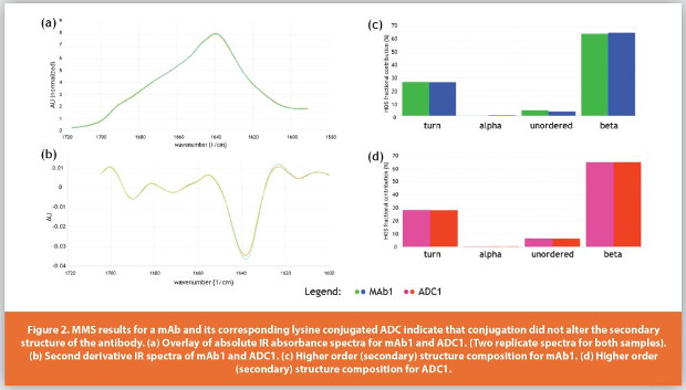
Figure 2 shows a first dataset for a native mAb and its corresponding lysine-based conjugated ADC. With this linking technology, the drug is attached to unmodified lysine residues across the mAb and the aim here was to confirm that the structure of the antibody had remained intact after being conjugated.
Absolute IR absorbance spectra for the Amide I band typically take the form of a single broad peak (Figure 2a); two sets of duplicate measurements are included in this plot, though barely visible, illustrating the excellent reproducibility of the data. Second derivative data (Figure 2b) brings resolution to the results unpacking the information in the absolute spectra and revealing characteristic bands at specific wavenumbers. Absorption features of the Amide 1 band are well correlated with shifts in hydrogen bonding and dipole-dipole interactions relating to α-helices, β-sheets, turns and other motifs of secondary structure so these data can be used to determine the fractional contribution of each element, using the instrument’s integrated software (see Figure 2c and 2d). The results indicate that around 65% of the secondary structure of both the native and conjugated mAb exists as β structure, a Figure that is in close agreement with literature values for this type of antibody;1,2 the fractional contributions of other major types secondary structure motifs are also similar. Even after conjugation, the structure of the mAb remains largely unaltered.
With cysteine-based conjugation technology, free sulfhydryls of disulfide bonds need to be made available via a process of reduction to enable attachment of the drug payload. When using engineered cysteine conjugation technology, the HOS of the antibody must therefore redevelop, post processing, to be closely similar to that of the original mAb. In a second study, a mAb and its corresponding cysteine-conjugated ADC were analyzed to assess parity between the native and conjugated antibody (data not shown). The results obtained were very similar to those for the lysine conjugated ADC, indicating that the secondary structure of the mAb and that of the ADC were comparable.
In the preceding tests, MMS is confirming consistency between samples - the absence of change - rather than detecting a change where one would be expected. To challenge this aspect of performance, MMS was used to assess the results of thermal stress tests, which are commonly applied to investigate the stability of biotherapeutics. In a first test, duplicate measurements of an ADC sample were carried out before and after storage at 50˚C. Though minor differences are observable in the absolute and second derivative spectra (not shown), the fractional contribution data indicate that under these conditions there is no significant change in secondary structure (Figure 3a), with a similarity score for the control and stressed samples based on an area of overlap analysis of > 99% . However, SEC data for the same samples indicate that the stressed sample contains ~22 – 25% aggregates (see Figure 3b). An explanatory hypothesis for this result is that the applied temperature, though sufficiently high to impact the quaternary structure and aggregation state of the mAb, does not change the more fundamental secondary structure of the antibody/ADC.

In the second temperature stress experiment, the sample was held at 70°C for 20 minutes. Analyses carried out in triplicate for the control and stressed samples (Figure 3c) exhibit excellent reproducibility and show a marked shift from parallel to anti-parallel beta sheet structure.
The similarity score for the two samples was just 76%. IR is particularly sensitive to the β-sheet structures that are prevalent in proteins (e.g. monoclonal antibodies) and to the changes in intermolecular β-sheet structure which accompany aggregate formation. These MMS results provide strong evidence for the formation of aggregates and are directly supported by the SEC data (Figure 3d) which shows that around 90% of the sample has aggregated.
Such tests demonstrate the ability of MMS to sensitively detect aggregation processes that impact secondary structure, with the precise resolution of beta sheet structure, a particularly useful capability. At the same time the MMS results highlight that in certain instances aggregation may leave the secondary structure of the protein unaltered. This too is valuable information when developing the comprehensive understanding of product behavior, most specifically aggregation behavior and mechanism, required to support development.
Alongside temperature, pH is one of the variables most commonly associated with structural change, so a comparable pH stress experiment was carried out by holding an ADC at pH 3 for three days. This produced similar results to the high temperature stress test (see Figure 4a) with a similarity score for the stressed relative to the control sample of just 78%. There was clear evidence of the shift from parallel to anti-parallel beta sheet structure associated with aggregation in the lower pH sample. SEC data again provided confirmation of aggregation under these conditions (data not shown).

In a further test, to assess the feasibility of using a new formulation that was almost 2 pH units lower than the current platform formulation, a different ADC exhibited much lower sensitivity to pH difference. The new formulation enables better management of certain product attributes but is only viable if HOS can be maintained under the somewhat different conditions. Here, MMS proved an efficient way to check structural integrity in the new formulation, the results showing that the secondary structure of the ADC remains comparable despite the lower pH, with equivalent fractional contributions of all elements of secondary structure (see Figure 4b).

In a final test, MMS data were directly compared with those produced by conventional FTIR (see Figure 5). The results showed good agreement between the two absolute and second derivative spectra, and the assignments of fractional content for the major α-helix and parallel β-sheet structures, as were also comparable. Though there were some notable differences with respect to the other structural element assignments (e.g. turns, random coil). These are attributable to the different software used by the two methods for curve fitting and peak assignments.
Conclusion
IR spectroscopy is widely recognized as a valuable tool for characterizing the secondary structure of proteins, which in turn is important during the development of ADCs, and biotherapeutics in general. The introduction of MMS provides a reliable, sensitive, high throughput technique for measurement of the secondary structure of biotherapeutics and offers a workflow regime and sample requirements that make it well-suited to screening studies early in the development process.
These studies show that MMS can efficiently support the development of ADCs by helping to answer important questions relating to the effects of conjugation on the secondary structure of the mAb, the stability of the product with respect to thermal and pH stress, and formulation screening studies. By generating this information more easily than traditional techniques, such as FTIR or CD, MMS has the potential to enable the more prevalent use of IR spectroscopy in biopharmaceutical development and to play a valuable role in the development of important novel therapeutics such as ADCs.
References
- Levitt M, Greer J. Automatic identifi cation of secondary structure in globular proteins. J. Mol. Biol. 1977;114(2):181-239.
- Byler DM, Susi H. Examination of secondary structure of proteins by deconvoluted FTIR spectra. Biopolymers 1986;25(3):469-487
Author Biography
Karan K. Shah is a Principal Development Associate in the Analytical and Pharmaceutical Sciences department at ImmunoGen, Inc. He received his M.S. in Pharmaceutical Sciences from Northeastern University and is an analytical chemist with several years of experience in the biopharmaceutical industry. His R&D experience includes formulation development as well as analysis and characterization of antibodies and antibody drug conjugates (ADCs). He has expertise in method development for characterization of antibodies and ADCs using a broad spectrum of analytical and biophysical techniques and in the application of these techniques to support CMC development.