Abstract
Mass spectrometry imaging (MSI) is a new and evolving analytical platform for molecular distribution studies, enabling multiplexed analyses of a wide variety of chemical species without their a priori selection. MSI can be used to distinguish diseased from healthy tissues using molecular marker-based characterization studies. In addition, it has been used effectively to determine the location of drugs and their metabolites in tissue samples collected from animal model systems. Not surprisingly, MSI is becoming a vital technology for biomedical and pharmaceutical research.
Text
 Mass spectrometry imaging (MSI) is well-suited for the analysis of a wide variety of samples including heterogeneous and complex biological tissues. But what is it? MSI combines the chemical characterization abilities of mass spectrometry but does so while providing spatial information. The most common approach is to acquire a mass spectrum by impinging a beam of light or ions at a specific sample location; by moving the beam stepwise across a tissue sample and acquiring a mass spectrum at each location, an image is created, one pixel at a time. As the beam is rastered across the sample, the image is created. Acquiring large images can be a slow process as literally tens of thousands of locations are interrogated. Compared to most optical imaging approaches, the spatial resolution is not good, but the chemical information from each location is high, especially given the outstanding performance capabilities of modern mass spectrometers. Because the chemical detail does not require labeling analytes of interest, unexpected details often emerge such as unique chemical forms or surprising metabolites of drugs. The goal of this article is to provide an overview of several MSI applications; for specifics about the instrumentation and technology, the reader is referred elsewhere [1, 2].
Perhaps the two most commonly used types of MSI are secondary ion mass spectrometry (SIMS) [3-7] and matrix-assisted laser desorption/ionization (MALDI) MS [8-13]. SIMS is well-suited for high spatial resolution imaging (below one micron), including subcellular imaging, of low molecular weight species (<500 Da). MALDI MS is typically applied to higher molecular weight species (>500 Da) within tissue sections of larger areas at a spatial resolution greater than 10 μm. Both approaches are still evolving in terms of their figures of merit, but already many biomedically relevant studies have been conducted [14]. As MSI matures into a more finely-developed analytical technique, it will become a routine tool in the pharmaceutical and medical fields for discovery and diagnostic applications.
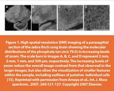
SIMS imaging has been used to provide insight into the distributions of lower molecular weight chemical species including vitamin E, cholesterol, and lipids [6, 11, 15-18]. Monroe et al. [16] reported the subcellular distribution of vitamin E within single neurons isolated from Aplysia californica. Vitamin E actively concentrates along the soma-neurite junction; its distribution is difficult to measure due to its integration into the phospholipid bilayer, which prevents it from being easily labeled using immunohistochemistry. Amaya et al. [15, 19] used SIMS imaging to generate individual >10 million pixel images of several chemical species including phosphate, arachadonic acid, and palmitic acid from zebra finch parasagittal brain sections. Figure 1 illustrates the ability of SIMS imaging to create chemical distribution maps ranging from molecular distributions across a tissue region, with individual pixels being on the scale of individual larger cells. SIMS imaging has the potential to enable fundamental studies where high spatial resolution is required in order to answer important biomedical questions.
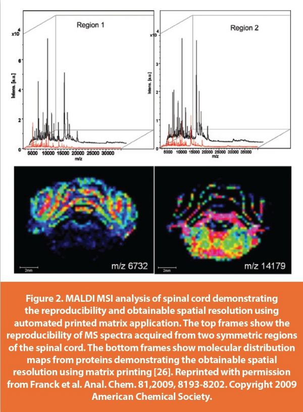
Whereas SIMS excels in the lower mass range, MALDI MS is well-suited for analysis of higher molecular weight species such as intact lipids [10, 12, 13], peptides [11, 20-22], and proteins [9, 13, 23]. MALDI MSI performance has been improved with respect to sample reproducibility and throughput—two factors that are paramount to its use in large-scale biomedical investigations. Sample preparation plays a key role in determining the quality of the resultant images [24, 25]. Tissue sections prepared for MALDI MSI analysis are coated with a homogeneous layer of matrix crystals prior to analysis. The homogeneity of this layer determines, in part, the quality of the images that are produced. Rather than manually spray coating samples with matrix, automated matrix application systems that use either spray or discrete spot printing are now available. Franck et al. [26] investigated the reproducibility of mass spectra quality when printing matrix for the analysis of low molecular weight proteins. Figure 2 from their work illustrates the high reproducibility and high mass spectra quality that can be obtained with automated matrix printing systems; the two molecular distribution maps exemplify the image quality that is achievable using this matrix application technique. These new matrix deposition systems will play a significant role in standardizing a protocol for biomedical studies by ensuring reproducibility.

Biomedical studies can demand a higher throughput than proof of principal demonstrations of a technology. Perhaps not surprisingly, acquiring tens of thousands of mass spectra in order to generate each image can be time consuming and often takes several hours.
Fortunately, the throughput of MALDI MSI analysis has steadily improved. A novel imaging technique recently developed by Spraggins and Caprioli [27] allows for continuous raster MALDI MSI. A spatial resolution of 100 μm is achievable in 10 min for a sample area of 185 mm2, which represents an improvement of 100-fold from previous imaging speeds. This novel imaging strategy results in a dramatic increase in throughput, thereby making MSI more feasible for routine biomedical analyses.
In addition to imaging endogenous species, MSI can also be used to track the localization and fate of xenobiotics, including pharmaceutical drugs, without developing a specific label for each substance. In addition, a drug’s metabolic products can be monitored simultaneously owing to the multiplexed detection capabilities inherent to MS. As an example, Khatib-Shahidi et al. [28] followed the fate of olanzapine within rats at different time points after administration. In order to perform this study, they made use of the multiple reaction monitoring capabilities offered by their hybrid MALDI MS system to enhance the sensitivity and selectivity of the analysis [28]. Images collected 2 h after administration, with the tmax as determined by previous autoradiography studies [29], are shown in Figure 3. Interestingly, olanzapine was found to localize throughout the body while its metabolites localized to the bladder, kidney, and liver. This approach can be expanded to monitor the localization of multiple drugs in a single organism, which could provide a method to study drug interactions. Studies of this nature yield invaluable information that can be used to help further drug development and biomedical research.
MSI is capable of enabling discovery-type experiments without a priori knowledge of the analytes of interest. It has developed into a reliable technique for laboratory analyses of a range of tissue samples, including molecular species that are difficult to image using other methods. As the technical figures of merit of MSI improve, especially spatial resolution and speed, concurrently with the development of more robust sampling protocols, we expect MSI to be used for a wider range of pharmaceutical studies. Accordingly, MSI technologies promise to be at the forefront of biological and medical discoveries during the coming years.
References
- Rubakhin, S.S. and Sweedler, J.V., eds. Mass Spectrometry Imaging: Principles and Protocols. First ed. Methods Mol. Biol. Vol. 656. 2010, Humana Press: New York, NY.
- Setou, M., ed. Imaging Mass Spectrometry: Protocols for Mass Microscopy. 2010, Springer: New York, NY.
- Boxer, S.G., Kraft, M.L., and Weber, P.K., Advances in imaging secondary ion mass spectrometry for biological samples. Annu. Rev. Biophys., 2009, 38:53-74.
- Fletcher, J.S., Lockyer, N.P., Vaidyanathan, S., and Vickerman, J.C., TOF-SIMS 3D biomolecular imaging of Xenopus laevis oocytes using buckminsterfullerene (C60) primary ions. Anal. Chem., 2007, 79:2199-2206.
- Kurczy, M.E., Piehowski, P.D., Van Bell, C.T., Heien, M.L., Winograd, N., and Ewing, A.G., Mass spectrometry imaging of mating Tetrahymena show that changes in cell morphology regulate lipid domain formation. Proc. Nat. Acad. Sci. U.S.A., 2010, 107:2751-2756.
- McDonnell, L.A., Piersma, S.R., Altelaar, A.F.M., Mize, T.H., Luxembourg, S.L., Verhaert, P.D.E.M., van Minnen, J., and Heeren, R.M.A., Subcellular imaging mass spectrometry of brain tissue. J. Mass Spectrom., 2005, 40:160-168.
- Nygren, H., Börner, K., Hagenhoff, B., Malmberg, P., and Månsson, J.-E., Localization of cholesterol, phosphocholine and galactosylceramide in rat cerebellar cortex with imaging TOF-SIMS equipped with a bismuth cluster ion source. Biochim. Biophys. Acta, 2005, 1737:102-110.
- Cornett, D.S., Reyzer, M.L., Chaurand, P., and Caprioli, R.M., MALDI imaging mass spectrometry: molecular snapshots of biochemical systems. Nat. Meth., 2007, 4:828- 833.
- Groseclose, M.R., Malin, A., William, M.H., and Richard, M.C., Identification of proteins directly from tissue: in situ tryptic digestions coupled with imaging mass spectrometry. J. Mass Spectrom., 2007, 42:254-262.
- Hankin, J.A., Barkley, R.M., and Murphy, R.C., Sublimation as a method of matrix application for mass spectrometric imaging. J. Am. Soc. Mass Spectrom., 2007, 18:1646-1652.
- Monroe, E.B., Annangudi, S.P., Hatcher, N.G., Gutstein, H.B., Rubakhin, S.S., and Sweedler, J.V., SIMS and MALDI MS imaging of the spinal cord. Proteomics, 2008, 8:3746–3754.
- Murphy, R.C., Hankin, J.A., and Barkley, R.M., Imaging of lipid species by MALDI mass spectrometry. J. Lipid Res., 2009, 50:S317-322.
- Tucker, K.R., Serebryannyy, L.A., Zimmerman, T.A., Rubakhin, S.S., and Sweedler, J.V., The modified-bead stretched sample method: development and application to MALDI-MS imaging of protein localization in the spinal cord. Chem. Sci., 2011, 2:785-795.
- Rubakhin, S.S., Jurchen, J.C., Monroe, E.B., and Sweedler, J.V., Imaging mass spectrometry: fundamentals and applications to drug discovery. Drug Discov. Today, 2005, 10:823-37.
- Amaya, K.R., Monroe, E.B., Sweedler, J.V., and Clayton, D.F., Lipid imaging in the zebra finch brain with secondary ion mass spectrometry. Int. J. Mass spectrom., 2007, 260:121-127
- Monroe, E.B., Jurchen, J.C., Lee, J., Rubakhin, S.S., and Sweedler, J.V., Vitamin E imaging and localization in the neuronal membrane. J. Am. Chem. Soc., 2005, 127:12152-12153
- Lanekoff, I., Kurczy, M.E., Adams, K.L., Malm, J., Karlsson, R., Sjövall, P., and Ewing, A.G., An in situ fracture device to image lipids in single cells using ToF-SIMS. Surf. Interface Anal., 2011, 43:257-260
- Mas, S., Touboul, D., Brunelle, A., Aragoncillo, P., Egido, J., Laprevote, O., and Vivanco, F., Lipid cartography of atherosclerotic plaque by cluster-TOF-SIMS imaging. Analyst, 2007, 132:24-26
- Amaya, K.R., Sweedler, J.V., and Clayton, D.F., Small molecule analysis and imaging of fatty acids in the zebra finch song system using time-of-flight-secondary ion mass spectrometry. J. Neurochem., 2011, 118:499-511
- Zimmerman, T.A., Rubakhin, S.S., Romanova, E.V., Tucker, K.R., and Sweedler, J.V., MALDI mass spectrometric imaging using the stretched sample method to reveal neuropeptide distributions in Aplysia nervous tissue. Anal. Chem., 2009, 81:9402-9409
- Chen, R., Hui, L., Sturm, R.M., and Li, L., Three dimensional mapping of neuropeptides and lipids in crustacean brain by mass spectral imaging. J. Am. Soc. Mass Spectrom., 2009, 20:1068–1077
- DeKeyser, S.S., Kutz-Naber, K.K., Schmidt, J.J., Barrett-Wilt, G.A., and Li, L., Imaging mass spectrometry of neuropeptides in decapod crustacean neuronal tissues. J. Proteome Res., 2007, 6:1782-1791
- Seeley, E.H. and Caprioli, R.M., Molecular imaging of proteins in tissues by mass spectrometry. Proc. Nat. Acad. Sci. U.S.A., 2008, 105:18126-18131
- Heeren, R.M.A., Kükrer-Kaletas, B., Taban, I.M., MacAleese, L., and McDonnell, L.A., Quality of surface: the influence of sample preparation on MS-based biomolecular tissue imaging with MALDI-MS and (ME-)SIMS. Appl. Surf. Sci., 2008, 255:1289-1297
- Zimmerman, T.A., Monroe, E.B., Tucker, K.R., Rubakhin, S.S., and Sweedler, J.V., Chapter 13 Imaging of cells and tissues with mass spectrometry: adding chemical information to imaging. Methods Cell Biol., 2008, 89:361-390
- Franck, J., Arafah, K., Barnes, A., Wisztorski, M., Salzet, M., and Fournier, I., Improving tissue preparation for matrix-assisted laser desorption ionization mass spectrometry imaging. Part 1: Using microspotting. Anal. Chem., 2009, 81:8193-8202
- Spraggins, J. and Caprioli, R., High-speed MALDI-TOF imaging mass spectrometry: rapid ion image acquisition and considerations for next generation instrumentation. J. Am. Soc. Mass Spectrom., 2011, 22:1022-1031-1031
- Khatib-Shahidi, S., Andersson, M., Herman, J.L., Gillespie, T.A., and Caprioli, R.M., Direct molecular analysis of whole-body animal tissue sections by imaging MALDI mass spectrometry. Anal. Chem., 2006, 78:6448-56
- Chay, S.H. and Herman, J.L., Disposition of the novel anti-schizophrenic drug [14C]olanzapine in male Fischer 344 and female CD rats following single oral dose administration. Arzneimittelforschung., 1998, 48:446-454.
Author Biographies
Kevin Tucker is currently a Mass Spectrometry Specialist with the School of Chemical Sciences Mass Spectrometry Laboratory at the University of Illinois at Urbana-Champaign. Dr. Tucker received his Ph.D. in analytical chemistry in 2011 for the development of sample preparation methods for mass spectrometry imaging, including both MALDI-MSI and SIMS imaging.
Jonathan Sweedler is the James R. Eiszner Family Professor of Chemistry as well as of Director of the School of Chemical Sciences at the University of Illinois at Urbana-Champaign. His research emphasizes analytical neurochemistry. He develops new analytical technologies for small-volume peptidomics and metabolomics, including single cell mass spectrometry and mass spectrometry imaging. He is currently Editor-in-Chief of Analytical Chemistry.
This article was printed in the September/October 2012 Digital Edition of American Pharmaceutical Review. Copyright rests with the publisher. For more information about American Pharmaceutical Review and to read similar articles, visit www.americanpharmaceuticalreview.com and subscribe for free.