Bacterial Endotoxin Test (BET) users seek ways to overcome low endotoxin recovery (LER) from direct spikes into undiluted biologics. These studies have come about from Chen’s initial observation1 that Control Standard Endotoxin (CSE) spikes, when placed into undiluted biologics, buffers, and other constituents often cannot be recovered. By changing the endotoxin spike requirement from diluted product to undiluted product, users will unsurprisingly encounter test interference; what is unexpected from Chen’s LER finding is that recovery sometimes cannot be improved by dilution. This is indicative of a still undefined binding phenomenon. Chen originally dismissed protein binding as a mechanism of LER as placebo with no protein of the same formulation also exhibited a low endotoxin spike recovery problem. However, we should not assume that the binding phenomenon with protein and without protein demonstrate the same mechanism of spike loss.

Figure 1. Interactive causal factors in the loss of endotoxin spike into undiluted product. Factors contributing to spike loss include areas where (1) protein binding dominates, (2) the phenomenon of spike disassociation to monomers dominates and (3) a mixture of factors 1 and 2 occurs. The bottom portion represents no binding/ masking and the top represents difficulties in spike recovery
This article will discuss a couple of different phenomena to explain LER observations (see Figure 1); they may be dependent on both the protein concentration and the protein charge (anionic versus cationic) as well as surfactant and excipient effects:
- High protein concentration or highly cationic proteins may demonstrate endotoxin-protein binding as Petsch et al observed in 1998 (discussed next section) [1, 2]
- Low and no protein content, and anionic (negativelycharged) protein samples may undergo spike disassociation via polysorbate dispersal and be exacerbated by citrate and/or phosphate
- The interplay of the two phenomenon may confound clear-cut mechanism determinations for a given protein formulation
Granted the description of “low” and “high” protein concentration as well as the cationic or anionic nature of specific proteins have to be established. Users must allow for the initial ambiguity of not knowing which of a variety of effects poor spike recovery may arise. Every subpar spike recovery from undiluted product should not be called LER; particularly if it can be overcome using traditional BET tools or tools associated with protein unmasking. It is an expectation that in trying to recover spike from undiluted product (now called “LER studies”) users will likely see test interference of many kinds and only those particularly difficult scenarios containing polysorbate and citrate/phosphate loss of spike over time should technically referred to as “LER”. The other option – that LER stands for anything adverse that can happen to an undiluted spike – seems too vague.
Protein binding Component
In 1998, Petsch, Deckwer and Anspach [1, 2] showed that protein binding of endotoxin is in fact an expected part of human antibody interaction with endotoxin2 in vitro when they mixed IgG and an E. coli filtrate. The study shows that they were able to quantify the extreme binding and to unbind or de-mask the protein solution using a protease (proteinase K) to recover an amount equivalent to the initial spike (see Table 1). A few observations from the Petsch paper appear most significant as related to LER:
Table 1. Condensed table from Petsch, Deckwer, and Anspach’s Tables 1 and 4 [1, 2]
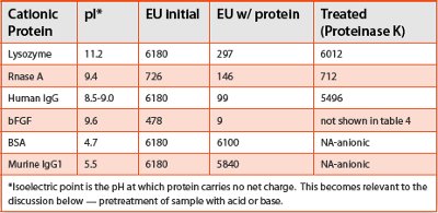
- There was no polysorbate or citrate involved in these solutions
- Anionic proteins (BSA and mouse IgG1) presented no difficulty in detecting added endotoxin
- Very large amounts of endotoxin (~6,000 EU) were bound by the proteins
- A very small amount of protein was used in their study – each solution was 1 mg/mL – some mAbs are 10-100 times more concentrated
- IgG is arguably a model protein that represents monoclonal antibodies
Feasibility studies performed here utilizing a couple of proteases, including the one Petsch used, reveal the ability to greatly improve direct spike recoveries in some cases from biologics containing protein, polysorbate and citrate. This would not likely be the case if protein binding were not at least one component of spike loss for some molecules. However, the use of proteases leaves something to be desired from a cGMP vantage as proteases often retain a (low) level of endotoxin (they are cationic proteins also) that is difficult to fully remove (Petsch admits as much) and may be expensive to use on a routine basis.
Since monoclonal antibodies are the product of a specific technology shared amongst industry participants, it stands to reason that in developing tools for de-masking similar molecules (IgG variants), then the tools developed should find some widespread utility amongst industry participants, yet to date it seems that user solutions have been highly formulation-dependant. As shown in Figure 1, protein binding is likely dependent upon both the concentration of the protein [3] and the associated overall charge, whereas the spike dissociation effect has been demonstrated by Chen and others to vary with regard to non-protein formulation constituents.
Dissociation of endotoxin spikes into monomers
The biologically active form of endotoxin has been shown to exist as aggregates (micelles) [4]3. The irreversible dispersal of aggregates via polysorbate has been hypothesized to be a potential cause of reduced recovery of direct spike. However, it should be noted that if the dissociation of endotoxin spikes into monomers were the only basis of the LER phenomenon, then we could not (a) explain the utility of a protease in de-masking a given biologics solution, (b) explain the utility of a basic pre-treatment with a similar effect (to be described) and (c) integrate Petsch’s data into current spike loss recovery observations. It seems more reasonable to believe that the LER phenomenon is split into at least a couple of different, and possibly interacting, mechanisms of action. If this is true, as shown in Figure 1, then the presence of polysorbate may turn out to be more anecdotal than necessary to the protein component of the LER effect for some biologics, particularly at either high protein content and/or with highly cationic proteins
A German biotech company focused on phage-ligand and recombinant factor C technology has conducted a great deal of work developing endotoxin de-masking solutions that may be necessary to employ for biologics, buffers, placebo, etc., see Figure 2. The company has demonstrated methods that de-mask variou
The use of naturally occurring endotoxin (NOE) from user-grown organisms that are harvested, killed aqueous solutions reminiscent of those used by Bowers and Tran [7] have been proven to help overcome the dissociation of spike effect, however, Johannes Reich presented data at the 2014 PDA Conference in Berlin demonstrating that masking occurs with NOE just as it does with CSE (though it may be delayed) and in a formulation-dependant manner that includes polysorbate and citrate just as Chen had first described. Of course, every NOE is likely different (the pros and cons of its usage in a nutshell).
Method Development aids to utilizing the “screening test”
As previously discussed [8], the concept of “screening” biologics to (a) determine if a given formulation may or may not be subject to LER, LER-like effects, or protein binding and (b) develop methods to overcome the loss of direct spike has been on-going since it was first introduced. This section is an overview of developmental tools that may add utility including: (1) the use of a proteins’ isoelectric point (pI), (2) use of RSE, (3) use of staggered development testing, (4) protease pretreatment, (5) de minimis pretreatment and transfers and (6) a combination treatment consisting of pH pretreatment and protease. Each section contains potential “next steps” to explore (bullet points).

Figure 3. Different proteins have different isoelectric points. Manipulation of the sample pH can remove a proteins’ charge.
Isoelectric point manipulation
The protein binding phenomenon is encountered and overcome in the biologics manufacturing environment during drug production and purification. Proteins are purified by running repeatedly through chromatography columns containing various immobilized ionic ligands. Various immobilized positively-charged ligands including Protein A (bind mAbs for subsequent elution), as well as a wide variety of cationic proteins [2] have been used to bind negatively charged endotoxin molecules (due to Lipid A dual phosphate groups) and other contaminants as they pass through. The theory of such removal methods revolves around the isoelectric point (pI) for any given protein (See Figures 3 and 4). Petsch showed the pI to be around 8 to 9.5 for the IgG used in Table 1 [1, 2]. Therefore, at pH 8 to 9.5 depending upon the specific protein, samples below pH 8 (commonly parenteral drugs are formulated around pH 6 - 7) can be treated with base to neutralize their charge and thereby free up endotoxin in solution. The key for BET is to obtain a strong enough basic pH without so much alkaline (or for so long a pretreatment time) that it becomes depyrogenating to the sample. The use of an acid or base pretreatment (depending on the protein’s pI) may find broad utility where precautions are taken against potential depyrogenation.
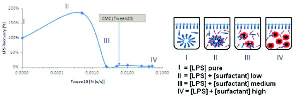
Figure 2. Effect of spike aggregation properties on LAL recovery in surfactant. I and II show low spike dissociation and high assay recovery. III and IV show high dissociation of spike with associated loss of endotoxin recovery [6].
Petsch and Anspach say that with basic proteins, “electrostatic interactions can be seen as the main driving force” but also point out that protein-endotoxin binding of “even acidic proteins (pI<7) are known, taking place also at low ionic strength” [1, 2]. The manipulation of a proteins’ charge via the isoelectric point is the same effect used in ion-exchange chromatography to remove contaminants from monoclonals in production environments; however, in production, the ligands (either cationic or anionic) are bound to the column or resin (immobilized). Testing previously documented [8] with mAb B, where very low initial recoveries were greatly improved upon using traditional diluents but remained declining into day 7, down to the low 70% range have been improved upon out to seven days using a method derived from isoelectric point principles.
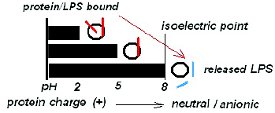
Figure 4. pH pretreatment. Isoelectric point (pI): pH where charge on a given protein becomes neutral. Illustrated for a cationic protein where pI = ~8.
Next steps:
- Titrating lower both the ph of the basic pretreatment solution and the volume needed to reach the isoelectric point (to reduce potential depyrogenation concerns)
- Better buffering of the post-basic treatment solution to decrease recovery variability
- Exactly defining the time sample is in the basic pretreatment solution
- Checking potential depyrogenation using a control endotoxin containing no filler
Reference standard endotoxin
The use of RSE may find utility as the potency of the lyophilized vial prior to reconstitution is 10,000 EU/vial. Method development using larger spike values may be preferable for determining trends in development treatments rather than measuring more minute levels associate with CSE (after dilution). When reasonable recovery is obtained it can be tweaked downward or switched over to CSE. RSE is not inexpensive yet the time spent on such studies can also quickly add up and developing methods faster obviously provides cost savings
Next steps:
- Testing using lower endotoxin spike levels for tests developed using high RSE concentrations
- Switching RSE development studies to CSE
Staggered Development testing
Spiking vials and allowing them to sit a couple of days and then working with the 48 or 72 hour samples (and beyond) to develop methods seems preferable to gaining spike recovery at zero and 24 hours with a method only to watch it decline into the coming days. Developing a method that works for the more difficult to recover sample spikes (aged) first can help speed and minimize the test development process. After all, the spike is still in the vial and the appropriate treatment will release it.
Protease pretreatment
As we have seen, Petsch et al. used proteinase K to free bound endotoxin from IgG samples (Table 1), however, the use of protease seems suboptimal from a GMP vantage as proteases often contain residual endotoxin. They can also be expensive. Purifying proteases may make them a viable option and they can be effective when combined in equal volume with a sample aliquot prior to dilution.
Next steps:
- Using protease pretreatment to confirm if the mechanism of spike loss involves protein binding for a given sample
- Developing purer protease solutions or alternatively titrating the amount used to very small levels (to treat correspondingly small sample aliquots similarly as in the basic pretreatment)
- Note that protease also may address hydrophobic endotoxinprotein binding that is not due to change
De minimis pretreatment and transfers
A minimalist approach to pretreatment with acid, base, or protease can serve to great effect. A 0.1 mL aliquot of a sample combined with a 0.1 mL aliquot of treatment solution for a determined time and temperature in a small depyrogenated glass tube can later be brought up to total volume of 1 mL (add 0.8 mL), with diluent added to the same tube to make a 1:10 dilution and then followed up with dilution to the desired test concentration. This is a small thing but cuts down on the number of transfers and thus the potential for spike loss.
Next steps:
- Trying to effect a change in the sample aliquot prior to further dilution and many treatment types can be tried in less time and effort than by using traditional pretreatments.
- Adding 0.1 mL sample to 0.1 mL pretreatment followed by the addition of 0.8 mL of diluent with mixing followed by another addition of 9 mL diluent (same tube) and continued mixing (1:100) carries the above concept further.
Combining ph pretreatment with protease
A couple of proprietary products are available that take advantage of both a pH/ isoelectric point pretreatment combined with a subsequent protease treatment. Combining the effects of both while minimizing the adverse effects of each provide powerful tools for products where other, simpler, solutions have failed.
Next steps:
- Simpler is better, but having a complete (simple to complex) repertoire of development tools sooner rather than later as deadlines approach is ideal.
Summary
Users continue to make strides in addressing LER-related issues from various perspectives. Here some potentially overlapping and interactive causation factors (Figure 1) and methods to overcome such factors have been explored. The matching of diff erent mechanisms with corresponding tools on a molecule-to-molecule basis is critical to overcoming what appear to be multiple factors contributing to the loss of spike recovery from direct spikes into undiluted drug product. Perhaps the most useful and interesting phenomenon going forward is the concept of utilizing a protein’s isoelectric point (pI) to neutralize the charge on a cationic protein, thereby potentially releasing endotoxin bound to protein to make it available for BET assay. Indeed, the underlying theory enjoys universal use in manufacturing environments historically.
A simple screening approach using a standardized endotoxin (CSE or RSE) seems flexible, able to test various proposed mechanisms of spike loss, and preferable to the tedious calibration of a widely variable, nonstandardized endotoxin (study-to-study and user-to-user), provided it proves widely applicable to many different biologics (different proteins, with various charges, concentrations, and/or formulations), and to nonprotein containing buffers and other constituents. Ideally, users want evidence that binding or spike dissociation has been overcome in the test matrix by the method used, not to obtain results that depend upon the use of a specially tailored endotoxin spike solution
Author Biography
Kevin Williams, currently at Hospira Inc., has 30 years’ experience in the Pharmaceutical industry specializing in endotoxin testing and control. He has written extensively on the subject of LAL technology including authoring/editing the book “Endotoxins” (Informa Healthcare, 2007).
References
- Petsch, Deckwer, Anspach. “Proteinase K digestion of proteins improves detection of bacterial endotoxins by the Limulus Amebocyte Lysate assay: application for endotoxin removal from cationic proteins”. Analytical Biochemistry, 259, pp. 42-47, 1998.
- Petsch and Anspach. “Endotoxin Removal from Protein Solutions”. Journal of Biotechnology, 76 pp. 97-119, 2000.
- Li and Luo. “Protein concentration effect of protein-lipopolysaccharide (LPS) binding and removal”. Biotechnology Letters, Vol. 19, No. 2, pp. 135-138, Feb. 1997.
- Mueller et al. “Aggregates are the biologically active units of endotoxin”. Jour. Biological Chem, Vol. 279, No. 25, pp. 26307-26313, June 18 2004.
- EndoLISA®: a novel and reliable method for endotoxin detection, Application Notes, Nature Methods, Oct. 2011.
- www.hyglos.de
- Bowers, K. and Tran, L. “Creation of an In-house Naturally Occurring Endotoxin Preparation for Use in Endotoxin Spiking Studies and LAL Sample Hold Time Analysis”. American Pharmaceutical Review, Vol. 14 Issue 6, 2011.
- Williams, K. “Endotoxin Test Concerns of Biologics”. American Pharmaceutical Review. Vol. 16 Issue 6 Endotoxin Detection Supplement, 2013.