Abstract
This review provides some insights into the chromatographic tools available for the detailed characterization of monoclonal antibodies (mAbs), and antibody drug conjugates (ADCs), since they represent an emerging class of therapeutic agents currently being developed by most of the pharmaceutical companies. The latest trends in size-exclusion chromatography (SEC), ion exchange chromatography (IEX), hydrophobic interaction chromatography (HIC) and reversed phase liquid chromatography (RPLC) will be critically exposed.
Current developments of biopharmaceuticals
There is currently a revolution occurring in the pharmaceutical field. With the quest for more targeted therapies and more clinically efficacious drugs, biopharmaceutical companies are increasing their efforts to develop more and more biologics.
Monoclonal antibodies (mAbs) are an emerging class of therapeutic agents under development in most pharmaceutical companies. These innovative molecules are used for multiple indications, including inflammatory disorders, autoimmune diseases, and cancer. In the last 30 years, there have been nearly 60 antibody products and derivatives approved by the EMA (European Medicines Agency) and the FDA (United States Food and Drug Administration), and the growth in approved mAbs in the last few years has been exponential. Today, more than 400 mAbs, primarily involving immunological and oncological targets, are under pre-clinical development and clinical trials, with an approval rate of approximately 20% compared with 5% for new chemical entities. Several characteristics of mAb therapy contribute to its success by improving the risk-benefit ratio. These characteristics include improved tolerance, high specificity, limited side effects and good efficacy. Numerous major first-generation mAbs will come off patent soon in Europe and the USA. Therefore, there are currently 35 biosimilar mAbs in clinical trials in Europe1 and two biosimilars of infliximab were recently approved by the EMA.2 Many companies are interested in producing copies or “generic” versions of commercial mAbs, but they are much more complicated to produce than small molecule generics.3
Because the curative potential of “naked” mAbs is rather limited, significant efforts have been devoted to the “arming” of mAbs, with bioactive payloads. The most interesting payloads are cytotoxic drugs, which display high potency toward cancer cells, but very little selectivity and high systemic toxicity. In this context, considerable efforts were invested in recent years to combine the desirable properties of mAbs (i.e. specificity and multi-functionality) with the cell killing activity of the most powerful cytotoxic drugs.4 Although the concept of antibody-drug conjugates (ADC) appears simple, its successful development was very recent and the first successful commercial ADC only appeared on the market in 2011. Today, the area of ADCs development is in rapid expansion with large investments from pharmaceutical companies.5
Requirements for mAbs and ADCs characterization
Because mAbs exhibit a high molecular complexity, they may be quite sensitive to changes in the manufacturing processes that can lead to considerable micro-heterogeneity in each individual chain. There are several common modifications leading to antibody charge variants and size variants on the peptide chains or variation of glycan profile. The combination of these micro-heterogeneity sources significantly increases the overall micro-heterogeneity in an entire IgG and should be critically evaluated because differences in impurities and/or degradation products can lead to serious health implications.6 In general, the identity, heterogeneity, impurity content, and activity of each new batch of mAbs should be thoroughly investigated before release and several important characteristics have to be considered.
The assessment of primary structure is generally one of the first characterization steps, since it allows confirming the theoretical molecular mass, including major post-translational modifications (PTMs).7 The size variants occurrence, including aggregates (larger components than the individual mAb) and incomplete formation of disulfide bridges (components smaller than the intact mAb) should also be evaluated since such variants are commonly observed in a mAb structure and can take place during production, formulation and storage of mAb product. As an example, aggregation of mAbs is an irreversible process that has a deleterious effect on both safety and efficacy, being a potential cause of enhanced adverse side effects and immunogenic responses.8 Charge variants are also commonly observed, and both acidic and basic species can exist, compared to the main isoform. Among the possible charge variants of the native mAb product, deamidation of asparagine, C-terminal lysine truncation and N-terminal pyroglutamation have been reported as important variants impacting the protein structure and function. Finally, the glycosylation pattern can strongly vary depending on the species employed for producing mAbs. As discussed elsewhere, the glycan profile should be deeply characterized since it can influence binding affinity, stability and safety of mAbs.8
In comparison with the “unconjugated” mAb, the ADCs present an increased level of complexity because the heterogeneity of the conjugation is superimposed on the variability associated with the initial mAb. Indeed, the conjugation technology results in an ADC product that is heterogeneous, with respect to both the loading of cytotoxic drug species and its distribution on the mAb. To better characterize these products and have sufficient information to support process and formulation development, routine lot-release and stability testing, several characteristics of ADCs have to be determined.9
Average drug to antibody ratio (DAR) corresponds to the average number of drug molecules that are conjugated per antibody molecule. This determines the amount of payload (number of cytotoxic drugs) that can be delivered to the tumor cell, and affects both the toxicity and safety of the ADC. Drug-load distribution corresponds to the distribution of drug linked forms (i.e., fraction of antibodies containing zero, one, two, three, …, n drugs). This characteristic is important since different forms may have different toxicological and pharmacological properties. The presence of size variants should also be assessed because the presence of high molecular size variants (aggregates) could modify the pharmacokinetics (faster clearance) and reduce drug exposure. Because cytotoxic drugs linked to antibody are lipophilic, they increase the chance of forming aggregates during manufacturing and storage. Finally, the amount of free (unconjugated) drug should also be precisely determined for an ADC product, as the free drug could be critical in terms of toxicity and safety. Residual quantities of unconjugated drug or drug-related impurities may remain in the final product as a result of incomplete removal by purification steps.
For mAbs or ADCs characterization, a panel of separation techniques based on both liquid chromatography and electrophoresis is often employed, but most of the current methods are only able to see the major structural differences. It is also important to note that mass spectrometry (MS) plays a pivotal role in the structural elucidation of mAbs but also ADCs because it offers an additional degree of separation by mass/charge ratio, greatly facilitating the characterization of variants.10
Ion exchange chromatography
Ion exchange chromatography (IEX) is a historical technique widely used for the characterization of therapeutic proteins and can be considered as a reference and powerful technique for the qualitative and quantitative evaluation of charge heterogeneity. Among the different IEX modes, cation exchange chromatography (CEX) is the most widely used for protein characterization.
As a classical mode of IEX, a linear salt-gradient is regularly applied for the elution. Proteins are eluted in order of increasing binding charge (correlates more or less with the isoelectric point (pI)) and equilibrium constant). Ion-exchange chromatofocusing represents a useful alternative to linear salt-gradient elution IEX, for separating protein isoforms with minor differences in the pI. Chromatofocusing is performed on an ion-exchange column employing a pH gradient that can be generated internally within the column.
Highly cross-linked non-porous poly(styrene–divinylbenzene) (PS/ DVB) particles are most frequently used in protein separations due to their pH stability (2 ≤ pH ≤ 12).
Moorhouse et al. were among the first ones to describe the potential of IEX for mAb characterization.11 Papain-digested mAb samples were successfully separated and the corresponding fragments were identified thanks to MS detection. A more recent study showed the applicability of a shallow pH gradient through CEX monolithic column and demonstrated relatively high resolution separation of mAb charge variants in three different biopharmaceuticals.12 Zhang et al. presented a multiproduct charge sensitive separation method for 16 mAbs possessing pI-s between 6.2 and 9.4.13 The fast and efficient separation of cetuximab Fab (fragment of antigen-binding) and Fc (fragment cristallizable region) variants were recently reported in both salt and pH-gradient CEX mode.14,15Figure 1 shows the separation of these variants by using a linear salt-gradient.
 Figure 1. Separation of the charge variants of papain digested Cetuximab by salt-gradient based CEX. Peak 1 and 2 correspond to the main Fc and main Fab fragments. Adapted from Ref [14], with permission.
A systematic study compared the possibilities of IEX modes and showed that these approaches are a cost- and time saving alternative to classical protein analysis methods (e.g. gel electrophoresis). The authors predicted that in the next step, further biologicals, e.g. antibodies, will be analyzed and quantified mostly with IEX and RPLC in the native as well as in its denatured form, respectively.16
Size exclusion chromatography
Beside IEX, size exclusion chromatography (SEC) is another historical technique widely employed for the qualitative and quantitative evaluation of protein aggregates.17 The main advantage of this approach is the mild mobile phase conditions that permit the characterization of proteins with minimal impact on the conformational structure and local environment.
SEC separates biomolecules according to their hydrodynamic radius. The stationary phase consists of spherical porous particles with a carefully controlled pore size, through which the biomolecules diffuse based on their molecular size difference using an aqueous buffer as the mobile phase.
During recent years, there have been a number of advances in SEC that improve the quantity of information that can be gained from a single injection. Among them, the use of shorter (e.g. 15 cm) and narrower (4.6 mm i.d.) columns packed with smaller-sized particle (below 3 μm) to improve the throughput and resolution seems to be a new trend, but care should be taken to avoid the risk of shear degradation caused by high pressure (>400 bar).18
Various detectors may be multiplexed in SEC, including refractive index (RI), ultraviolet (UV), multi-angle laser light scattering (MALLS) and viscometer (IV), for extensive characterization of protein samples.19
A high-throughput SEC method was recently reported, that was able to differentiate candidate recombinant human monoclonal IgG1 and IgG2 antibodies based on their propensity to form aggregates when subjected to agitation stress.20 Wätzig et al. showed a 15-min SEC separation of IgG1 antibody aggregates and demonstrated the precision and repeatability of monomer and aggregate quantitation.16 By using state-of-the-art column technology and UHPLC instrumentation, it was demonstrated that the analysis time can be reduced to less than 3 minutes.18 Figure 2 shows the fast SEC separation of mAb aggregates.
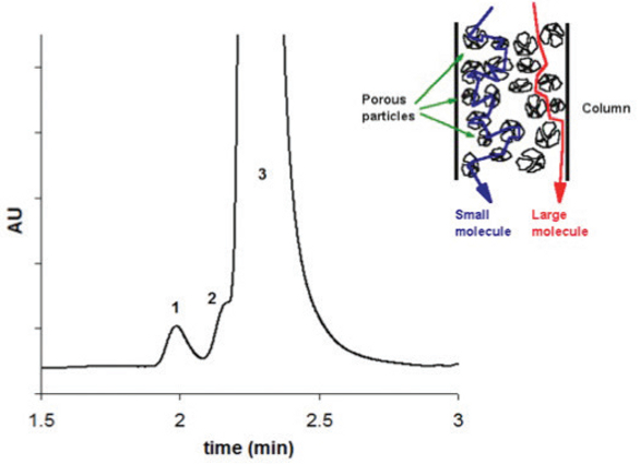 Figure 2. Fast separation of mAb aggregates by SEC. Peak 1, 2 and 3 correspond to high molecular weight species (HMW), dimer and monomer mAb, respectively. Adapted from Ref [18], with permission.
Similar to what is commonly performed for intact mAbs, the determination of ADC is carried out using SEC. However, regular SEC mobile phase often provides poor peak shape of ADC and unacceptable resolution between aggregates and monomeric ADC products.21 This result could probably be explained by non-specific interactions between the hydrophobic cytotoxic drugs and the surface of the stationary phase. To solve this problem and improve peak shape, various organic modifiers were added to the SEC mobile phase.22,23
Hydrophobic interaction chromatography
Hydrophobic interaction chromatography (HIC) is often used for the purification of proteins. Recently, the main applications for HIC becomes the estimation of DAR of ADCs.
HIC takes advantage of the hydrophobicity of proteins promoting its separation on the basis of hydrophobic interactions between immobilized hydrophobic ligands and non-polar regions on the surface of proteins.24 The adsorption increases with high salt concentration in the mobile phase and the elution is achieved by decreasing the salt concentration of the eluent. In HIC, the structural damage to the biomolecules is minimal and its biological activity is maintained.
One of the most important quality attributes of an ADC is the average number of drugs that are conjugated as well as the drug loading. HIC analysis allows both the characterization of the distribution of druglinked species and the determination of average DAR.25 Conjugation of the drug-linker to the antibody increases the hydrophobicity, therefore HIC is an appropriate tool to separate the different DAR species of ADCs. The HIC profile of IgG2 ADC is shown in Figure 3.
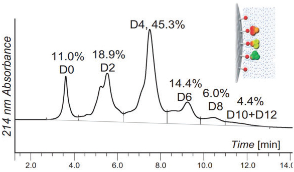 Figure 3. HIC separation of IgG2 ADC species. D0, D2, D4, D6, D8, D10 and D12 correspond to different drug conjugations from unconjugated (D0) to 12 conjugations (D12). Adapted from Ref [25], with permission.
The DAR of a commercial ADC, namely Brentuximab Vedotin (cystein linked IgG1) was recently assessed by offline HIC Native MS. Each individual HIC peak was collected, buffer exchanged, and analyzed by native MS. Native MS allowed to confirm mass homogeneity of each HIC fraction as no mixtures of different drug loading stoichiometries were observed within one HIC fraction.26
Recently, HIC columns packed with 2.5 and 3 μm particles and possessing 2.1 mm i.d. were released. This confirms the potential of high resolution HIC separations and shows that the current trend in HIC is similar to other chromatographic modes (miniaturization and increase of resolving power).
Reversed phase liquid chromatography
The main benefits of reversed phase liquid chromatography (RPLC) over any other LC modes are its high resolving power and compatibility with MS. In RPLC, solute retention is predominantly mediated through the hydrophobic interactions between the non-polar amino acid residues of the proteins and the bonded n-alkyl ligands of the stationary phase. The superior robustness of RPLC makes it well suited for use in a routine environment.27
The use of ultra high pressure liquid chromatography (UHPLC) systems and columns packed with highly efficient sub-2 μm particles has become popular for the separation of small molecules very soon after its commercial introduction in 2004. In 2006, the first RPLC material packed with widepore (300 Å) 1.7 μm particles was introduced and opened new avenues for protein analysis.28 Since then, several generations of superficially porous wide pore materials and organic monoliths have also been commercialized and make possible to perform very fast or high-resolution protein separations.28
The complete characterization of an intact mAb or ADC with RPLC is difficult to achieve. Therefore, various enzymes, such as pepsin, papain and IdeS are often used to obtain relatively large fragments and facilitate the investigation of their micro-heterogeneity. This limited proteolysis approach – or “middle down” – is mostly used recently for the RPLC characterization of mAbs.29 The reduction of disulfide bonds is also commonly used to produce two light chains (LC) and two HCs fragments of 25 and 50 kDa, respectively. To prepare only 25 kDa fragments, the reduced mAb (HC and LC) can be further digested with papain. In most cases, the limited proteolysis approach enables to determine the heterogeneity of mAb related to the different parts of the molecule.
Outstanding separations within relatively short analysis times (typically between six and ten minutes) were demonstrated on rituximab, bevacuzimab and panitumumab employing gradients between 30 and 40% acetonitrile, temperatures above 70°C and 0.1% TFA as ionpairing additive.30 As example, the RPLC separation of the variants of Bevacizumab’s Fc and Fab fragments can be seen in Figure 4.
 Figure 4. RP UHPLC separation of Bevacizumab Fc and Fab fragments. Peaks: 1–3: pre-Fc peaks, 4: Fc, 5,6: post-Fc peaks, 7–9: pre-Fab peaks, 10: Fab, 11–13: post-Fab peaks. Adapted from Ref [30], with permission.
The main application of LC-MS in the analysis of therapeutic proteins is the identification of the protein primary structure and impurities/degradation products. Nowadays, peptide mapping is performed under RPLC-ESI/MS (electrospray ionization) conditions. LC-MS based peptide mapping of mAbs typically allows covering over 98% of the sequence and confirming identity.27 Implementing tandem MS systems allows obtaining fragmentation data on the peptides and further enhancing the confidence of peptide identity, sequence and modification sites.27 As example, UHPLC was also combined with QqTOF/MS (quadrupole-quadrupole-time-of-flight) in a detailed study of mAb deamidation to achieve chromatographic resolution of the deamidated products, while maintaining relatively short analysis time.31
Conclusion
As illustrated in this review, the characterization of biopharmaceuticals is challenging and numerous analytical strategies have to be combined to have a global view on the microheterogeneity of mAbs or ADCs. IEX, SEC and HIC are historical orthogonal techniques offering a high selectivity for separating charge variants, size variants and hydrophobicity variants, respectively, from the main protein isoform. However, due to the significant developments in MS in the last few years, RPLC is gaining in importance, despite a lower resolving power than IEX, SEC and HIC. Except chromatographic techniques, it is also important to mention that electrophoretic as well as spectrophotometric approaches are also widely used for the characterization of biomolecules.
References
- Schneider CK, Vleminckx C, Gravanis I, Ehmann F, Trouvin JH, Weise M, Thirstrup S. Setting the stage for biosimilar monoclonal antibodies. Nat. Biotech. 2012;30:1179-1185.
- Beck A, Reichert JM. Approval of the first biosimilar antibodies in Europe: a major landmark for the biopharmaceutical industry. MAbs. 2013;5:621-623.
- Beck A, Debaene F, Diemer H, Wagner-Rousset E, Colas O, Dorsselaer AV, Cianférani S. Cutting-edge mass spectrometry characterization of originator, biosimilar and biobetter antibodies. J Mass Spectrom. 2015;50:285-289.
- Deonarain MP, Yahioglu G, Stamati I, Marklew J. Emerging formats for next-generation antibody drug conjugates. Expert Opin Drug Discov. 2015;10:463-481.
- Beck A, Reichert JM. Antibody-drug conjugates: present and future. MAbs. 2014;6:15-17.
- Fekete S, Gassner AL, Rudaz S, Schappler J, Guillarme D. Analytical strategies for the characterization of therapeutic monoclonal antibodies. TrAC. 2013;42:74-83.
- Beck A, Diemer H, Ayoub D, Debaene F, Wagner-Rousset E, Carapito C, Van Dorsselaer A, Sanglier-Cianferani S. TrAC. 2013;48:81-95.
- Beck A, Wagner-Rousset E, Ayoub D, Van Dorsselaer A, Sanglier-Cianferani S. Characterization of therapeutic antibodies and related products. Anal. Chem. 2012;85:715-736.
- Wakankar A, Chen Y, Gokarn Y, Jacobson FS. Analytical methods for physicochemical characterization of antibody drug conjugates. mAbs. 2011;3:161-172.
- Alvarez M, Tremintin G, Wang J, Eng M, Kao YH, Jeong J, Ling VT, Borisov OV. On-line characterization of monoclonal antibody variants by liquid chromatography-mass spectrometry operating in a two-dimensional format. Anal. Biochem. 2011;419:17-25.
- Moorhouse KG, Nashabeh W, Deveney J, Bjork NS, Mulkerrin MG, Ryskamp T. Validation of an HPLC method for the analysis of the charge heterogeneity of the recombinant monoclonal antibody IDEC-C2B8 after papain digestion. J. Pharm. Biomed. Anal. 1997;16:593-603.
- Talebi M, Nordbog A, Gaspar A, Lacher NA, Wang Q, He XZ, Haddad PR, Hilder EF. Charge heterogeneity profiling of monoclonal antibodies using low ionic strength ion-exchange chromatography and well-controlled pH gradients on monolithic columns. J. Chromatogr. A. 2013;1317:148-154.
- Zhang L, Patapoff T, Farnan D, Zhang B. Improving pH gradient cation-exchange chromatography of monoclonal antibodies by controlling ionic strength. J. Chromatogr. A. 2013;1272:56-64.
- Fekete S, Beck A, Fekete J, Guillarme D. Method development for the separation of monoclonal antibody charge variants in cation exchange chromatography, Part I: salt gradient approach. J. Pharm. Biomed. Anal. 2015;102:33-44.
- Fekete S, Beck A, Fekete J, Guillarme D. Method development for the separation of monoclonal antibody charge variants in cation exchange chromatography, Part II: pH gradient approach. J. Pharm. Biomed. Anal. 2015;102:282-289.
- Grotefend S, Kaminski L, Wroblewitz S, Deeb SE, Kuhn N, Reichl S, Limberger M, Watt S, Watzig H. Protein quantitation using various modes of high performance liquid chromatography. J. Pharm. Biomed. Anal. 2012;71:127-138.
- Fekete S, Beck A, Veuthey JL, Guillarme D, Theory and practice of size exclusion chromatography for the analysis of protein aggregates. J. Pharm. Biomed. Anal. 2014;101:161-173.
- Fekete S, Ganzler K, Guillarme D. Critical evaluation of fast size exclusion chromatographic separations of protein aggregates, applying sub-2 μm particles. J. Pharm. Biomed. Anal. 2013;78-79:141-149.
- Hong P, Koza S, Bouvier ESP. A review, size exclusion chromatography for the analysis of protein biotherapeutics and their aggregates. J. Liq. Chrom. Rel. Techn. 2012;35:2923-2950.
- Woodard J, Lau H, Latypov RF. Nondenaturing size-exclusion chromatography-mass spectrometry to measure stress-induced aggregation in a complex mixture of monoclonal antibodies. Anal. Chem. 2013;85:6429−6436.
- King HD, Dubowchik GM, Mastalerz H, Willner D, Hofstead SJ, Firestone RA, Lasch SJ, Trail PA. Monoclonal antibody conjugates of doxorubicin prepared with branch peptide linkers: inhibition of aggregation by methoxytriethileneglicol chains. J. Med. Chem. 2002;45: 4336-4343.
- Hollander I, Kunz A, Hamann PR. Selection of reaction additives used in the preparation of monomeric antibody-calicheamicin conjugates. Bioconjug Chem. 2008;19:358-361.
- Quiles S, Raisch KP, Sanford LL, Bonner JA, Safavy A. Synthesis and preliminary biological evaluation of high-drug-load paclitaxel-antibody conjugates for tumor-targeted chemotherapy. J. Med. Chem. 2010;53:586-594.
- Queiroz JA, Tomaz CT, Cabral JMS. Hydrophobic interaction chromatography of proteins. J. Biotech. 2001;87:143–159.
- Wiggins B, Shin LL, Yamaguchi H, Ratnaswamy G. Characterization of cysteine-linked conjugation profiles of immunoglobulin G1 and immunoglobulin G2 antibody-drug conjugates. Pharm. Drug Deliv. Pharm. Tech. 2015;104:1362-1372.
- Debaene F, Boeuf A, Wagner-Rousset E, Colas O, Ayoub D, Corvaïa N, Van Dorsselaer A, Beck A, Cianférani S. Innovative native MS methodologies for antibody drug conjugate characterization: high resolution native MS and IM-MS for average DAR and DAR distribution assessment. Anal. Chem. 2014;86:10674-10683.
- Sandra K, Vandenheede I, Sandra P. Modern chromatographic and mass spectrometric techniques for protein biopharmaceutical characterization. J. Chromatogr. A. 2014;1335:81-103.
- Fekete S, Guillarme D. Ultra-high-performance liquid chromatography for the characterization of therapeutic proteins. TrAC 2014;63:76-84.
- Fekete S, Gassner AL, Rudaz S, Schappler J, Guillarme D. Analytical strategies for the characterization of therapeutic monoclonal antibodies. TrAC 2013;42:74-83.
- Fekete S, Rudaz S, Fekete J, Guillarme D. Analysis of recombinant monoclonal antibodies by RPLC: Toward a generic method development approach. J. Pharm. Biomed. Anal. 2012;70:158–168.
- Sinha S, Zhang L, Duan S, Williams TD, Vlasak J, Ionescu R, Topp EM. Effect of protein structure on deamidation rate in the Fc fragment of an IgG1 monoclonal antibody. Protein Sci. 2009;18:1573-1584.
Author Biographies
Dr. Szabolcs Fekete holds a PhD degree in analytical chemistry from the Technical University of Budapest. He worked at the analytical R&D department of Gedeon Richter Plc for 10 years. Since 2011, he is working at the University of Geneva, Switzerland. He contributed 60+ journal articles and book chapters. His main interests include HPLC, column technology, pharmaceutical and protein analysis.
Dr. Alain Beck is Senior Director, Antibody/ADC Physico-Chemistry and member of the board of directors of the CIPF. He contributed to the R&D of anticancer mAbs in collaboration with Merck and Abbvie, vaccines and peptides. He is inventor on 16 patents, author of near 140 publications and reports and associate editor of mAbs.
Dr. Davy Guillarme holds a PhD degree in analytical chemistry from the University of Lyon, France. He is senior lecturer at the University of Geneva in Switzerland. He authored 140 journal articles related to pharmaceutical analysis. His expertise includes HPLC, UHPLC, HILIC, LC-MS, SFC, analysis of proteins and mAbs. He is an editorial advisory board member of several journals including American Pharmaceutical Review.