Introduction
Classical microbiology techniques used in the quality control (QC) of food, cosmetics and pharmaceutical products as well as those used to monitor manufacturing environments are based on the growth of the potential microbial contaminants in liquid or solid nutrient media. These classic techniques usually require lengthy incubation times, typically on the order of days. Currently, there are a variety of rapid microbiological technologies commercially available that promise to significantly reduce these testing times and the associated delays in the release of manufactured products. Reduced testing times also offer increased capacity within existing manufacturing facilities, minimize required in-process inventory and faster monitoring and better control and understanding of the manufacturing process.
We are currently evaluating a number of alternative microbiological platforms and identifying the best match between QC requirements and the capabilities of a particular rapid assay. Detection of microbial adenosine triphosphate (ATP) via bioluminescence assays is one of the most familiar and robust rapid microbiological techniques used in the field of rapid QC. Adenosine triphosphate is an essential element of all living cells, serving as both the prime energy carrier and a regulator of enzyme activity within the cell (1). The bioluminescence reaction makes use of the firefly luciferase enzyme, which catalyzes the hydrolysis of ATP molecules and the release of a quantum of 564 nm yellow light for each molecule of the luciferin substrate oxidized (2). This reaction (see below) takes place in the presence of D-luciferin, and is dependent on the presence of oxygen and magnesium. Light produced from the luciferin-luciferase reaction can be measured in relative light units (RLUs) by a luminometer. Generally, the RLU measurements show a good correlation to the number of ATP molecules processed and thus the number of microbial cells present in the assay.

Intensive poultry production is required to meet an increasing worldwide demand for animal proteins. In the poultry industry, product quality is influenced by factors such as increased hatchability, reduced mortality and/or unmarketable birds, improved growth rate, better feed conversion and body composition (e.g., fat vs. muscle). Hatchery environments are usually hot and humid and therefore ideal for the proliferation of bacteria and molds. Newly hatched chicks rely on underdeveloped immune systems to protect them from infection. Therefore, a crucial factor to assure success in this industry is the implementation of effective vaccination programs to prevent and control the spread of disease (3). Vaccine use in poultry production usually translates into an increased production yield. Although the animal health industry has developed a variety of vaccines for the prevention of poultry diseases, they are not always administered to the birds in ways that ensure effective and consistent results. For instance, post-hatch vaccinations and field boosters can be very inconsistent resulting in inadequate protection. More recently, the development and commercial availability of automated in ovo injection technology has dramatically improved both the consistency and reliability of vaccination by inoculating chicks while they are still within the egg. The in ovo injection devices help provide an early immune response by vaccinating eggs prior to hatching with the added benefit of reducing bird stress. This automated system punches a tiny hole into the egg shell and then lowers a needle through the hole which delivers the therapeutic product safely and accurately to the embryo. This usually occurs on the 18th day of the bird’s total 21 day incubation period.
The use of automated chicken egg vaccine injectors by large commercial hatcheries has increased dramatically during the last decade. Concomitantly, there is a need to assure faster release of newly manufactured devices to meet the increased demand. One of the final steps in the release of the new devices to the marketplace involves a biochallenge test. The test is performed by QC to verify the effectiveness of the instrument sanitization procedure and is applied to all new injector systems prior to shipment from manufacturing to customers. The test involves an intentional contamination (i.e., challenge with E. coli) of the vaccine pathway followed by a sanitization cycle of this pathway and a subsequent microbiological assay to determine the effectiveness of the cycle. This study describes the evaluation of an ATP bioluminescence based rapid microbiology assay to monitor the effectiveness of a sanitization process after an introduced microbial challenge as an element of the quality control of a chicken egg vaccine injector device.
Results and Discussion
Monitoring the Effectiveness of Vaccine Pathway Sanitization Following an E. coli Challenge

Figure 1 - Chicken egg vaccine delivery system.
An example of a commercially available chicken egg injector system is shown in Figure 1. Some of these systems can deliver vaccines in ovo to chick embryos and are capable of injecting over 60,000 eggs per hour (depending on specific tray configurations). Since these devices are designed to operate in a relatively open, non sterile environment, such as those found in a commercial hatchery, it is imperative to apply a good sanitization procedure. To that effect, and under routine conditions, the system tested throughout our studies runs a sanitizing flush cycle of the whole vaccine pathway between each injection cycle. In addition, needles are individually sanitized after each injection. This significantly minimizes contamination carry-over when compared to manual in ovo or post-hatch injections. To verify that the sanitization procedure is working properly in each newly manufactured device, an E. coli challenge, a sanitization cycle, and a QC microbiological assay was executed as described in the following section.
The Traditional Microbiological Procedure
A typical routine microbiological assay, which is based on traditional plate count, takes up to 72 hours to be completed. It involves the following steps: After cleaning and sanitizing the vaccine pathway, non-challenge control TSA plate samples are secured. Utilizing a specific E. coli culture, a bio-challenge is introduced to the vaccine pathway. After a second cleaning and sanitizing cycle, TSA plate samples are taken as validation criteria for efficacy of the procedure. The total time of growing the E. coli culture. collecting all critical samples of this challenge model, data recording, and report issuing is approximately 96 hours.
The traditional plate count procedure acceptance criteria calls for ≤ 1 Colony Forming Unit (CFU) of E. coli cells per mL of collected sample. This criteria is in accordance with a QC requirement to achieve a bacterial reduction of 5 logs or greater. It is important to point out that the volume of sample collected for analysis under this traditional procedure was in the order of milliliters because of the small volume used to inoculate each plate. In addition, only a sample set of the total number of injection positions were included in the assay; therefore the total sample volume represented only a fraction of the device total pathway dead volume.
A Faster Microbiological Assay
Due to both the increased demand for the commercial product as well as a desire to continually improve the quality control of the released devices, the QC group sought an alternative test method comprising the following characteristics:
- More time and cost efficient than the traditional assay allowing an increased number of new instruments to be released to the marketplace in the same timeframe as allowed by the traditional method.
- Easy to integrate into current analytical lab structure.
- Improved assessment by providing more information and check points (e.g., detection of all viable microbial cells, including injured or stressed cells, and a larger sample volume representing the entire vaccine pathway of the device).
In order to achieve these desired attributes, the following modifications were incorporated into a new procedure:
- The total collected sample includes the entire dead volume of the vaccine pathway.
- A representative sample is collected from all injection positions to support analysis.
- The use of a membrane filtration based assay to allow concentration of a large sample volume to one filter for application of a suitable rapid assay.
Selection of the New Rapid Method
An ATP bioluminescence based rapid microbiology system was selected for this application. The selected system consists of a compact, portable luminometer, reagents and disposables. The assay is designed for the detection of microbial contamination based on ATP using the luciferin-luciferase substrate/enzyme system from the firefly, Photinus pyralis. The rapid microbiology system allows for the direct measurement of microbial contamination on a filter membrane. Filtration of a sample offers the possibility to increase the sample volume as well as concentrate microorganisms on the filter membrane surface. There are two reagents that are then added directly to the membrane filter in a sequential manner. The first reagent is an extractant that makes the intracellular ATP present in any microbial cell collected on the surface of the filter available for the luciferase reaction. The second reagent is the mixture of luciferin and luciferase needed for the bioluminescence reaction. The reaction initiates within a few seconds, with the resultant light emitted and measured by the portable luminometer.
Some of the characteristics considered in the rapid system selection process are summarized as follows:
- Membrane filtration based therefore allowing the use of a much larger sample (e.g, 1 Liter)
- Good references, familiarity and technical support
- Relatively low maintenance and cost
- Portable, requiring a minimum of laboratory bench space
- Relatively simple training and ease of use
- Easy verification of proper system operation prior to use
- Standard ATP kit allowing corroboration of calibration.
- Results (RLUs) that can be reasonably correlated to CFUs
- Minimal sample manipulation
- Validation experience
- Detection of viable cells, including those that could be injured or stressed as a result of the sanitization procedure (i.e., viable but non culturable), assuring a better QC.
Validation of the Rapid System

Figure 2 - Ruggedness and Linearity. RLU:ATP Correlation curves from 8 different experiments and 3 different operators.
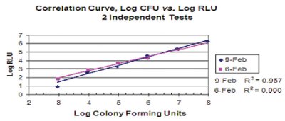
Figure 3 - Precision, Accuracy, and Linearity. Correlation of Colony forming Units to Relative Light Units for E. coli.
Table 1 - Uninoculated Control Analysis (Blanks). Blanks were processed under similar conditions as the assessment samples, but omitting the addition of either an ATP standard or microorganisms. Based on the background analysis shown in Figure 2, the acceptance criteria for a positive reading was defined as an RLU value of 400 (i.e., equivalent to approx 3x the value of the average blank reading)

Validation parameters were evaluated according to compendial references and the Parenteral Drug Association Technical Report #33 (4-6). The validation parameters obtained through this study were all satisfactory. Figures 2 and 3 and Table 1 illustrate the results obtained for critical parameters (e.g., ruggedness, linearity, accuracy, and precision).
Numerous correlation curves of E. coli and other microorganisms were run by us during the course of this study. A representative correlation curve is shown in Figure 3. The limit of detection for the bioluminescence based instrument was found to vary between 100 to 1,000 cells (as determined by correlated RLU values) for the assay run directly on membrane filters. This Limit of Detection (LOD) is in agreement with the results obtained by others (7). Based on this LOD value there was a need to adapt the rapid assay by increasing the volume of the collected sample. As mentioned previously, the acceptance criteria for the traditional test is ≤ 1 Colony Forming Unit (CFU) per mL of collected sample. By using a larger collection volume in the new rapid method we were able to have a sample large enough to represent the entire vaccine pathway of the injector, and to concentrate a sample large enough to be compatible with the LOD of the new rapid technology.. Regarding the LOD value, the acceptance criteria of the new rapid method was set at 1,000 CFU/L. In this way, any result above the limit of detection, is considered to be a failure of the sanitization process. The luminometer LOD was therefore adequate to meet a required 5 log reduction (or greater) of the applied bacterial challenge set by the QC inspection process. It is important to point out that the bioluminescence assay detects actual cells, but based on the correlation curves between the actual plate counts (CFUs) and bioluminescence units (RLUs), the acceptance criteria was defined as CFUs. In reality, the RLUs comprise cells able to generate colonies upon incubation on a solid media in addition to cells that could still be viable but unable to grow under the incubation conditions defined by the traditional methodology. Even though the probability that injured bacterial cells could survive the harsh sanitization process is remote, the new opportunity brought by the rapid assay to detect all type of viable cells represents a clear advantage by increasing our safety level and understanding of the effectiveness of the sanitization process.
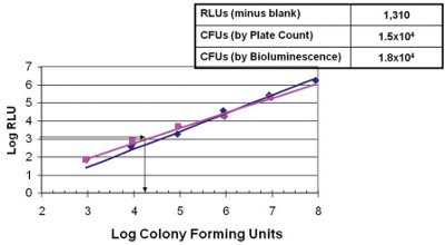
Figure 4 - Detection by bioluminescence assay of E. coli ATCC10536 cells recovered from a 1 Liter flush sample from a injector device spiked with approximately 104 bacterial cells.
To evaluate the performance of the new assay as well as test the sensitivity of the luminometer in a real case scenario, we conducted a challenge test of the vaccine pathway. The sanitized vaccine pathway was challenged with a number of E. coli cells (e.g., 104 cells) to a level just above the LOD of the new assay. The cells were held within the vaccine pathway for a set duration to allow adequate exposure and settling. The challenge media was cleared by flushing with 1 liter of sterile saline, recovered by membrane filtration and processed following the standard bioluminescence protocol. Figure 4 shows that the relative light units obtained with the luminometer gave an estimate of approx 1.8 x 10⁴ cells (or the equivalent CFUs) when interpolated in a standard RLU vs CFU correlation curve. The RLU value is in good agreement with the number of CFUs estimated by recovery of an aliquot of the same sample on TSA plates (1.5 x 10⁴). These results indicate that the rapid bioluminescence based assay constitutes a reliable alternative to the traditional method and that the sensitivity of this new rapid method is adequate to detect any potential residual bacterial presence above the five log reduction acceptance criteria for the sanitization process and release of the device.
A summary of the advantages of the new rapid method over the traditional assay
- A reduction in testing time and laboratory personnel time needed to perform the test. Conventional total time to results of 96 hours (using the traditional plate count method) was reduced to <48 hours. This represents a 50% time reduction in the total testing time.
- The bioluminescence based rapid method has a moderately reduced testing cost
- It reduced the amount of laboratory and incubator space needed over the traditional assay.
- Significantly decreased the time to release of the final product at the end of the manufacturing cycle.
- Larger sample size and better representation (i.e., entire vaccine pathway, including all needles).
- E. coli cells that might have survived the sanitization procedure and are injured or stressed have a greater probability of being detected.
- The selected luminometer is a portable instrument that permits near line processing of samples.
- Permits efficient use of troubleshooting time during assembly manufacturing process development.
Conclusions
A bioluminescence based rapid microbiological method was demonstrated to be as good as or better than a routine QC traditional microbiological release assay to determine sanitization effectiveness following a bio-challenge.
The results of this study show that the bioluminescence based rapid microbiology system is at a minimum reliable, sensitive, and equivalent to the standard or traditional plate count based test used to detect a required five log reduction in the level of E. coli cells inoculated during a bio-challenge step previous to the sanitization cycle of a vaccine chicken egg injector device. The assay is intended as the last step for the release of a newly manufactured injector device to the market. The implementation of the rapid method as a replacement to the traditional plate count based test allowed a significant decrease in manufacturing cycle time. APR
Acknowledgments
The authors wish to thank the following individuals: (1) Stewart Davenport, Senior Manager Technical Services, Pfizer Global Manufacturing, for his initial involvement and advise; (2) Corey Smith, Senior Manager, Pfizer Poultry Health, for his continued support to this project.
References
- Stanley, P.E. “A concise beginner’s guide to rapid microbiology using adenosine triphosphate (ATP) and luminescence” (1989) In ATP Luminescence: Rapid Methods in Microbiology, P. E. Stanley, B. J. McCarthy, and R. Smither, Eds., Blackwell Scientific Publications, Oxford, England, pp. 1-11.
- Lehninger, A.L. (1981) (Biochemistry, The Molecular Basis of Cell Structure and Function. Worth Publishers, Inc. New York.
- Marangon, S. and Busani, L. (2006). The use of vaccination in poultry production. Rev. Sci. Tech. Off. int. Epiz, 26 (1), 265-274.
- Parenteral Drug Association. (2000) Evaluation, validation and implementation of new microbiological testing methods. PDA J Pharm Sci & Technol; 54(3), suppl. TR33:2-9.
- United States Pharmacopeia. (2006) Chapter <1223> Validation of Alternative Microbiological Methods
- European Pharmacopoeia (2006) Chapter 5.1.6 – Alternative Methods for Control of Microbiological Quality
- Kramer, M., Suklje-Debeljak, H., and Kmetec, V. (2008). Preservative Efficacy Screening of Pharmaceutical Formulations using ATP Bioluminescence. Drug Dev and Industrial Pharmacy, 34, 547-557.
Claudio Denoya is a Research Fellow and Group Leader of the Microbiological Technology Assessment group in Pfizer Global R&D, responsible for evaluating and implementing rapid microbiological methods throughout the Pfizer organization. He is also an Adjunct Professor at the Department of Molecular and Cell Biology, Univ. of Connecticut. He led several other groups at Pfizer working on Streptomyces genetic engineering, Recombinant DNA, Eukaryotic Transfection & Expression, and Biologics & Microbiological Testing. He has authored over 200 patents, book chapters, journal articles and technical presentations. Dr. Denoya holds a Ph.D. in Biochemistry and Molecular Genetics of Animal Viruses, a M.S. in Biochemistry and Microbiology, and a B.S. in Clinical Biochemistry from the University of Buenos Aires. Readers may contact the author directly at claudio.d.denoya@pfizer.com
Edwin Dawson is a Biointegration Specialist for Pfizer Animal Health, located in Durham, North Carolina. Edwin is responsible for continuous internal and external customer support in the development, validation, and implementation of quality improvements to the in ovo vaccination process and clean-in-place operation of our commercial device. Edwin has more than 10 years of laboratory, manufacturing, and quality experience in the pharmaceutical industry and received his Bachelor of Science Degree in Biology from the University of North Carolina at Pembroke and his MBA from Indiana Wesleyan University.
Darwin Sessoms is a Biointegration Specialist that supports our validation department with In-house and Field device testing for new Pfizer in ovo equipment and components. He is also involved with the development, validation, and implementation of quality improvements to the in ovo vaccination process. Darwin has more than 10 years of Poultry health experience with a background in Field Services and Biological QC testing. Darwin has a Bachelor of Science Degree in Animal Science and a Master’s Degree in Animal Health Sciences from North Carolina Agricultural and Technical State University.
Michael Baumstein is an industrial microbiologist that has worked within the Pharmaceutical Industry for more than 17 years. Michael joined the Upjohn Company in 1992 after graduating with a Bachelors Degree in Microbiology from the University of Illinois – Urbana/Champaign. He worked within Quality Assurance and he has familiarity in all facets of microbiological monitoring of Aseptic Processing Operations and associated pharmaceutical utilities. He also has experience performing computer validation activities culminating in the development, management, and administration of an electronic environmental monitoring database system. Currently, Michael works within Pfizer’s Global Quality Operations Center (GQOC) - Microbiology/Aseptic Support group that provides microbiological technical support on a variety of topics for Pfizer manufacturing sites around the world and is a member of Pfizer’s Rapid Microbiological Methods Application Development team.
John Shabushnig is currently a Sr. Manager in Pfizer’s Global Quality Operations, responsible for providing microbiology and aseptic manufacturing technical support to manufacturing sites worldwide. John holds a B.S. in Chemistry from Carroll University and a Ph.D. in Analytical Chemistry from Indiana University. He has published and presented numerous papers on the subjects of spectroscopic analysis, process analytical technology (PAT) and visual inspection of pharmaceutical products. John is an active member of the Parenteral Drug Association (PDA), having recently served as Chair of the Board of Directors. He continues to serve on the Board of Directors, Executive Committee, Strategic Planning Committee and Science Advisory Board and as the leader of the Visual Inspection Interest Group. He also serves on the USP Parenteral Products Industrial Expert Committee (PPIEC) and the Ad hoc Committee on Visual Inspection and is a member of the American Chemical Society (ACS).