Introduction
X-ray crystallography has been employed in research and development of small molecules pharmaceuticals for many years. Small molecule active pharmaceutical ingredients (API) and excipients can crystallize as polymorphs which can have different solubilities and stabilities. Changes in solubility and stability can lead to changes in the bioavailability of the molecule [1]. Using X-ray crystallography in identifying the polymorphic form of a crystalline active pharmaceutical ingredient is well established in the literature [2]. A branch of X-ray crystallography, powder X-ray diffraction, has been used to “fingerprint” a specific solid form of the crystalline API, so that any solid form changes can be identified throughout product development and commercialization [3, 4]. For small molecules, crystalline form changes can have a large impact on the bioavailability of the molecule because of the changes in solubility. In biotherapeutic formulation development crystalline changes in the formulation matrix can have an impact on the stability of the formulated biologic. This article contains examples and considerations in the use of powder X-ray diffraction in biotherapeutic formulation development.

Figure 1 - Bragg diff raction
When X-ray radiation impacts the studied solid, in this case a powder, the lattice spacing of the molecules in the solid behaves as a diffraction grating. The diffracted beam has different phases because the distance travelled by the beam is dependent on the plane of reflection. The distance difference is illustrated in, Figure 1. where the distance A,B,C is less than D,E,F which causes the phase difference in the reflected beam. When the diffracted waves are in phase there is an increase in the intensity, which is referred to as constructive interference. When the diffracted waves are out of phase there is a cancelling of the intensity, which is referred to as destructive interference. The constructive and destructive interference can be measured as different intensities in the X-ray beam at given angles.

Figure 2 - Typical amorphous and crystalline diff raction patterns
The X-ray pattern can be used to confirm that the sample being analyzed is amorphous. Amorphous materials do not have long range order like crystalline materials and therefore the resulting diffraction pattern does not show the typical series of peaks associated with crystalline materials. The diffraction pattern of amorphous materials shows a broad “halo” with few or a single maxima, Figure 2. Although this “halo” pattern does not uniquely identify the material being studied it does confirm that the material is amorphous, which is critical knowledge needed for characterization.
While, powder X-ray diff raction is used to identify and monitor the crystalline solid form of the active pharmaceutical ingredient in a small molecule formulation, in the large molecule lyophilized formulations the excipients form an amorphous or crystalline matrix. The crystallinity of this matrix can change during manufacture or storage and needs to be monitored. The lyophilization process involves the freezing of the liquid formulation matrix to a temperature at which the ice nucleation occurs and the solution freezes. During the freezing process the protein in the solution is exposed to the stress of temperature changes as well as changes in concentration induced due to cryoconcentration [5]. The formulation components in a biotherapeutic formulation protect the protein from the freeze drying stress of cryoconcentration and temperature changes. Disaccharide sugars and polyols, typically mannitol, sucrose, and trehalose are used as cryoprotectants. Although the mechanism of the protection may vary between hydrogen bond disruption or preferential exclusion it is necessary to use the disaccharide sugars and polyols as a protectant to prevent aggregation and chemical degradation [6, 7].
It is common practice to include a bulking agent in the formulation to improve the physical appearance of the cake. The most common bulking agents are glycine and mannitol, both of which can be crystalline in the final lyophilized cake [8].
The pH of biotherapeutic formulations is often optimized and maintained by a buffer system to assure protein stability. Recrystallization of the buffer salts can occur because of a reduction in solubility when exposed to lower temperatures during lyophilization [9]. The high concentration carboxylic acid sodium salts have shown a propensity to crystallize under lyophilization conditions. The recrystallized carboxylic acid sodium salts have been found to have an effect on the stability of the protein in the lyophilized formulation as inferred from the structural changes in the protein secondary structure observed using FTIR measurements [10]. In summary, crystallization of the components in the lyophilized formulations can be induced by the reduction in temperature during the lyophilization cycle.
The crystallization of the formulation components does not necessarily lead to the same physical form as the starting material, because of the change in free energy conditions. The temperature, concentration, and localized water activity for a particular component have changed during and after the lyophilization process [11]. This change in the environment makes it ideal for a change in the final physical form of the formulation components. An excipient may crystallize or remain amorphous. If it crystallizes, it could crystallize possibly as a polymorph or a hydrate of the starting form. The ability to form a hydrate is now accessible due to the presence of water in the formulation during the lyophilization steps [12]. This is best illustrated by mannitol, which is known to exist in at least three polymorphic forms, an amorphous form, and a hemihydrate form [13-17]. All of these forms have been identified in various formulations illustrating the need to monitor the freeze dried product by powder X-ray diffraction as changes in the form of excipient may lead to degradation of the protein.
In general, vaccine formulations contain a buffer for pH control, the antigen, an adjuvant and a delivery vehicle [18]. The antigen is the species which is recognized by the immune system, and is usually a protein or polysaccharide. The delivery vehicle can combine the antigen in a virus like particle or it can be carrier for the antigen such as a liposome. Vaccine formulations have similar composition considerations to most biotherapeutic formulations. Sugars are added to provide cryoprotection and sodium chloride or similar salt can be added as a tonicity modifier and a formulation stabilizer. Vaccine formulations differ in that they contain an adjuvant to provide an increased immune response. Aluminum hydroxide and aluminum phosphate are the most widely used adjuvants in vaccine formulations. The recombinant antigen protein or polysaccharide is deposited on the surface of the aluminum salt adjuvant during the formulation process. The surface area and surface charge are important factors associated with the capacity of the adjuvant particle to adsorb the antigen [19]. The capacity of the adjuvant particle has been correlated to the increased immune response elicited by the administration of the vaccine [20].
An increase in the crystallinity of the adjuvant can lead to an increase in its propensity to aggregate in suspension. [19]. There are several proposed mechanisms of action as to the function of the adjuvant in the increased immune response [21]. It is believed that a smaller adjuvant particle is more effective in producing an immune response due to the ability of the smaller particle to be taken up by dendritic cells [22]. The crystallinity of the dried solids has been correlated to the change in particle size distribution toward larger particles or aggregates. The crystallinity of the adjuvant has also been found to impact the adsorption capacity [23]. An increase in the crystallinity is shown to decrease the adsorptive capacity. Thermal treatment can increase the relative crystallinity of aluminum hydroxide and it can also convert to a different polymorphic form [24]. Both of these changes in physical form have an impact on the stability of the vaccine components in the formulation and the biological effectiveness of the vaccine.
The changes in the physical form of the sugars in lyophilized formulations and the changes in degree of crystallinity of the adjuvant in vaccine formulations have an impact on some aspect of the stability of the protein and formulation. As discussed above it is important to know and monitor the physical form of the components in the formulation for a variety of reasons. Powder X-ray diffraction is ideally suited to monitor the development of crystallinity as well as changes in crystalline forms. In this paper, we describe different methods for powder X-ray diffractometry of a monoclonal antibody formulation, a DNA delivery system, and a vaccine formulation.
Materials and Methods
Powder X-ray diffraction was performed using a copper X-ray source maintained at 40 kV and 40 mA to provide Cu Kα1 (1.5406 Å ) and Cu Kα2 (1.54439 Å) radiation with an intensity weighted average of (Kαave) 1.54184 Å. An area detector or a scintillation detector were used for X-ray detection on the system. Data were collected on the area detector system with a source angle of 3.5° and a detector angle of 17° for 120 seconds. Data were collected on the scintillation counter system with a scan from 5 degrees 2θ to 35 degrees 2θ with a 0.2 step size. Powder X-ray diffractometry of the samples was performed without any further treatment such as hand grinding. In addition, the experiments were performed in specially fabricated sample holders (of a modified polymer) to protect them from the ambient moisture.
Dynamic vapor sorption was carried out on a high throughput water vapor sorption system. Two scans were done at 25°C from 0-90-0% RH with a step size of 10% RH and a change in mass versus a change in time (dm/dt) of 0.01.
Lyophilization was performed in a commercially available shelf freeze drying system. Typically a 0.5 mL fill in a 2 mL tubing glass vial and a fluorotec coated rubber closure were used. The samples were frozen at -50°C. Primary drying temperatures and times were dependent on the formulation. The vials were backfi lled with nitrogen to 800 mbar or 1000 mbar, stoppered and capped.
Silver Filter Sample Holders
Silver membrane fi lters 0.2um 47mm were used to isolate the aluminum phosphate suspension samples. The sample was isolated using vacuum fi ltration and then transferred directly to the powder X-ray instrument on the silver fi lter.
Hygroscopicity of Lyophilized Materials
The amorphous nature of the lyophilized formulations makes the samples hygroscopic by nature. The rate of moisture sorption could be of concern during handling of lyophilized samples that are predominately amorphous. A sample of a sucrose containing formulation was studied using dynamic vapor adsorption. A rate of adsorption of 0.8% per hour was observed for relative humidity of 30%, Figure 3. Moisture adsorption is known to cause recrystallization of the sugar in the lyophilized formulations [25]. To study the eff ectiveness of the domed sample holders, a lyophilized formulation containing sucrose was studied. The formulation was transferred to the domed sample holder in a nitrogen purged drybox. The diff raction pattern was then collected to show the sample was amorphous. The domed holder containing the sample was then placed into a 40°C/75%RH chamber for 30 minutes. The diff raction pattern was then collected again. The diff raction pattern showed no observable change. The same sample was then stored in the 40°C/75%RH chamber overnight. The diff raction pattern collected post 40°C/75%RH shows a peak pattern which matches that of crystalline sucrose, Figure 4. This shows that the domed sample holders isolate the sample from atmospheric moisture for a suffi cient time period to collect a representative diff raction pattern, but at time periods greater than 30 minutes and high humidity, the sample may not be isolated from moisture uptake. Additional work would be necessary to establish specifi c limits of time versus relative humidity for each studied sample because of the diff erent rates of adsorption for each sample.
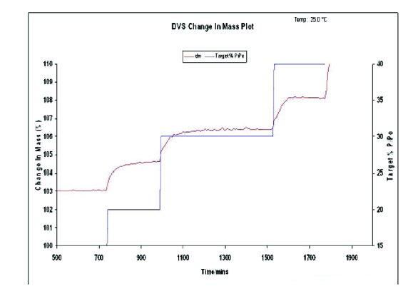
Figure 3 - Freeze dried cake rate of moisture adsorption.

Figure 4 -Domed sample holder moisture resistance
Results and Discussion
Case Study: mAb lyophilization
In the lyophilization process, the desired outcome is a freeze dried formulation which stabilizes the protein. Frequently the lyophilized formulation contains an amorphous domain which reduces the molecular mobility of the system and increases the stability of the protein [26, 27]. Because it is easy to distinguish an amorphous solid form from a crystalline solid form using powder X-ray, the technique is ideally suited to characterize freeze dried formulations. The overlay in Figure 5 shows the time equal zero point for three diff erent formulations. The formulations contain 85 or 125 mg/mL protein in several diff erent cryoprotectants, excipient combinations and a ratio of 1:2 polyol:disaccharide when the total amount of cryoprotectant was kept constant. The samples were buff ered using 20 mM buff er with a pH target of 5.8. The lyophilization conditions were similar to those previously discussed in the materials and methods section. The powder X-ray diff ractograms show that the formulations are all amorphous at the time equal zero point regardless of protein concentration or cryoprotectant combination. As part of the stability study the samples were stored at 5°C, 25°C, and 40°C. The samples were then studied at various time points to determine the stability of the amorphous matrix for 52 weeks. The time equal 23 weeks and the time equal 52 weeks overlay of select formulations at the studied temperatures are shown in Figure 6. The data indicates that there are no signifi cant changes in the solid form over 52 weeks at 5°C or 40°C. Establishing a consistent solid form over the time of the study is necessary so that changes in other chemical (oxidation, fragmentation) and physical (aggregation) assay can be attributed to the formulation and physicochemical properties and not due to a change in the solid form of the lyophile [28].

Figure 5 - Time Zero Freeze Dried Cake Diff ractogram

Figure 6 - Time 23 weeks and time 52 weeks stability sample diff ractograms
Case Study: Dihydrate excipient formation in delivery device
Powder X-ray diff raction is also well suited to identify the crystalline component of a complex mixture. Powder X-ray diff raction was used to identify a dihydrate excipient formation in a DNA formulation. The targeted formulation consists of a DNA plasmid precipitated with peptide, a chelating agent and an excipient on an inert carrier particle. The precipitation is carried out using a suitable antisolvent.
It is of interest to determine the solid form of the excipient deposited on the inert carrier particle to assure reproducibility in the fi nalproduct. It was also of concern that an ethanol or isopropanol solvate could form during the precipitation process. Because the excipient crystalline anhydrous and crystalline dihydrate forms are documented in the commercial single crystal libraries it is easy to compare the forms present in the process to the known forms. The starting material can be positively identifi ed as the excipient dihydrate by matching the collected raw material diff ractogram with the powder pattern predicted from the single crystal data Figure 7.
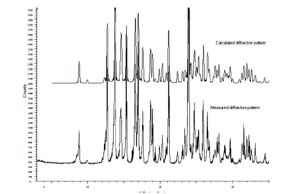
Figure 7 - Excipient calculated and measured diff raction patterns
The powder X-ray diff raction pattern of the formulated carrier particles are observed, however no other crystalline peaks are observed. In a separate experiment, the precipitation process was carried out in the absence of the carrier particles to produce the solid precipitate. The precipitate was then isolated on a silver fi lter and the powder pattern was collected. A typical amorphous halo was observed, indicating that no crystallization of any of the components had occurred.
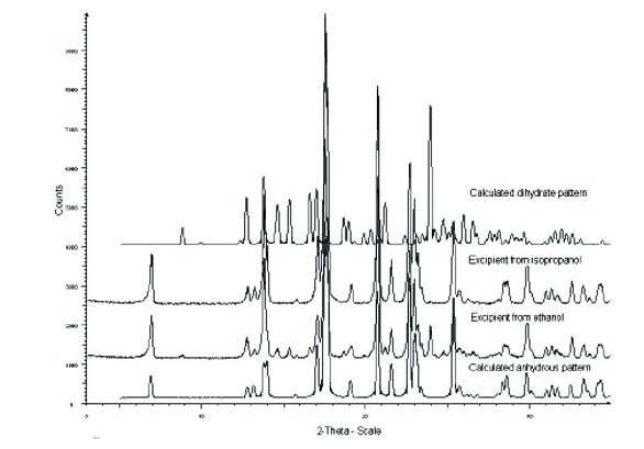
Figure 8 - Diff raction patterns of excipient isolated from ethanol and isopropanol
Our attempts to prepare solvates from slurries in ethanol and isopropanol were unsuccessful. Powder X-ray diff raction experiments revealed formation of anhydrous excipient in these slurries. No solvate formation was observed, but the powder X-ray pattern of the isolated solids from the slurry showed the formation of the anhydrous excipient in both ethanol and isopropanol slurries, Figure 8. It has been postulated that excipient is an effi cient stabilizer in protein formulations because of its hydration volume [29]. A change in the physical form from the amorphous form to a crystalline dihydrate, crystalline solvate, or crystalline anhydrate would change the hydration volume and consequently change the stabilizing eff ect of excipient. It is therefore important to identify the fi nal form of excipient to ensure that there is no change in the stability of the protected protein in the formulation.
Case Study: Vaccine-1
Vaccines, including recombinant subunit proteins, antigen conjugates (e.g. peptide or protein conjugates), are formulated with appropriate excipients and possibly an immunostimulant that may be deposited on the surface of an inorganic adjuvant. They are then delivered to the patient to elicit an immune response. The formulation can be complex, with multiple conjugates, diff erent immunostimulants and multiple stabilizing components.
Changes in the adjuvant physical structure can be assessed using powder X-ray diff raction data. The inorganic adjuvant can be isolated via fi ltration directly onto a silver fi lter. The silver fi lter is then placed into the powder X-ray diff ractometer and the diff raction pattern collected. The pattern is largely amorphous but shows small peaks indicative of some crystalline order, Figure 9. These peaks can be used to help diff erentiate the formulations. A series of 16 formulations, which contain diff erent salt to sugar ratios were lyophilized under identical conditions and studied using powder X-ray diff ractometry. Using the isolated inorganic adjuvant as a reference pattern, it is shown that undercertain conditions the amorphous lyophile can be observed, while in others the small peak identifi ed as the adjuvant can be observed, Figure 10. The adjuvant peak is observed only in formulations with a disaccharide at levels lower that 23 mg/mL.
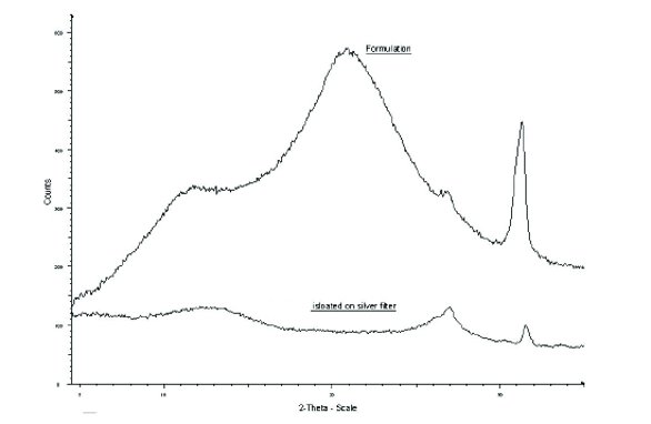
Figure 9 -Inorganic adjuvant and vaccine formulation powder X-ray diff raction patterns
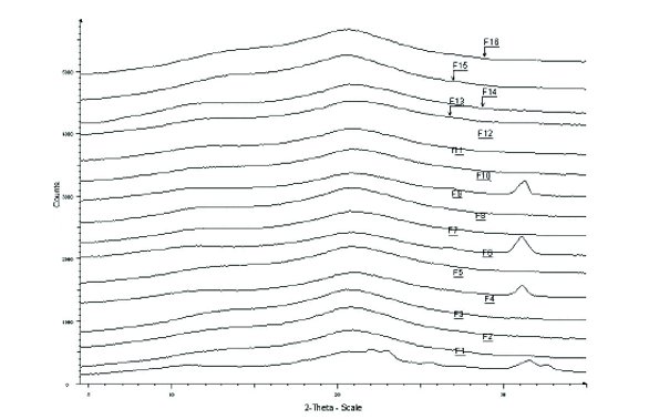
Figure 10 -Vaccine formulation diff raction patterns
Case Study: Vaccine-2
Powder X-ray diff raction was used to monitor the recrystallization of mannitol in freeze-dried formulations of conjugate vaccine. In this formulation study the formulation may contain a mixture of two proteins, combinations of disaccharides and bulking agents, mannitol, an immunostimulant and the inorganic adjuvant. Twelve formulations were studied, of these twelve, six were formulated with the inorganic adjuvant and six were formulated without the adjuvant. Powder X-ray diff ractometry data shows the presence of crystalline mannitol in the freeze-dried samples. The ratio of disaccharide to mannitol in the formulations is 1.2:1. The presence of the inorganic adjuvant or protein does not suppress the crystallization of the mannitol. The presence of the adjuvant and the protein together does not suppress the crystallization of the mannitol, however the formulation without the proteins and without the adjuvant resulted in the mannitol being in an amorphous form, Figure 11. The diff raction patterns are the same in all the formulations which show crystalline mannitol formation. The mannitol phase component in these formulations was identifi ed as a mixture of β- and δ- mannitol, Figure 12. There is evidence to show that the biologic activity of protein in a formulation with recrystallized mannitol is lower than that of the amorphous mannitol system [8, 30]. Therefore, it is important to monitor the physical form of mannitol in the formulation to maintain formulation consistency and biological activity.
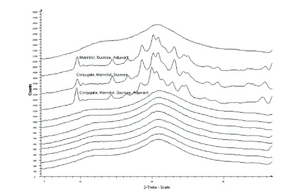
Figure 11 -Mannitol crystallization in formulation

Figure 12- Vaccine formulation compate
Conclusion
The use of powder X-ray diff raction is not necessarily restricted to small molecule research and development. It can be a useful tool to study the lyophilized formulations of large molecules, including monoclonal antibodies, virus like particles, and delivery device systems to just name a few. Powder X-ray diff raction can be used to confi rm the amorphous form of the starting lyophilized formulation and follow the physical form through the stability program. It can also be used to identify the crystalline components that appear in the formulation from the formulation process or stability study. The use of powder X-ray in biopharmaceutical development is a useful tool for monitoring the formulation, especially when the amorphous form is being used.
Crystallization of formulation components (cryoprotectants, buff er salts) can lead to a decrease in the stability and biological activity of proteins. Therefore, it is critical to monitor the physical form during processing, shipping, storage, and handling of freeze-dried protein formulations.
In small molecule development, the physical form of the active molecule plays an important role in the fi nal bioavailablility and effi cacy of the molecule. In biopharmaceutical development, the physical form of the formulation components plays an important role in the stability and therefore activity of the active moiety. Powder X-ray diffraction can be used to monitor the physical form in both development areas to assure that the final drug form is stable and has biological activity.
References
- Chemburkar, Sanjay R.; Bauer, John; Deming, Kris; Spiwek, Harry; Patel, Ketan; Morris, John; Henry, Rodger; Spanton, Stephen; Dziki, Walter; Porter, William; Quick, John; Bauer, Phil; Donaubauer, John; Narayanan, B. A.; Soldani, Mauro; Riley, Dave; McFarland, Kathyrn. Dealing with the Impact of Ritonavir Polymorphs on the Late Stages of Bulk Drug Process Development. Organic Process Research & Development (2000), 4(5), 413-417.
- Brittain, H Polymorphism in Pharmaceutical Solids Drugs an the Pharmaceutical Sciences Vol 95 Marcel Dekker New York 1999
- Bernstein, J Polymorphism in Molecular Crystals; Oxford University Press: New York, 2002.
- Byrn, S.R.; Preiffer, R.R.; Stowell, JG Solid State Chemistry of Drugs, 2nd ed; SSCI Inc.: West Lafayette, IN 1999
- Franks, F.. Freeze-drying: from empiricism to predictability. The significance of glass transitions. Developments in Biological Standardization (1992), 74(Biol. Prod. Freeze-Drying Formulation), 9-19.
- Shalaev, Evgenyi Y.; Johnson-Elton, Tiffany D.; Chang, Liuquan; Pikal, Michael J. Thermophysical properties of pharmaceutically compatible buffers at sub-zero temperatures: implications for freeze-drying. Pharmaceutical Research (2002), 19(2), 195-201.
- Arakawa, Tsutomu; Timasheff, Serge N. Preferential interactions of proteins with salts in concentrated solutions. Biochemistry (1982), 21(25), 6545-6552.
- Pyne, Abira; Chatterjee, Koustuv; Suryanarayanan, Raj. Solute crystallization in mannitol-glycine systems-implications on protein stabilization in freeze-dried formulations. Journal of Pharmaceutical Sciences (2003), 92(11), 2272-2283.
- Shalaev, Evgenyi Y.. The impact of buffer on processing and stability of freeze-dried dosage forms, Part 1: Solution freezing behavior. American Pharmaceutical Review (2005), 8(5), 80,82-87
- Izutsu, S. Kadoya, C. Yomota, T. Kawanishi, E. Yonemochi and K. Terada (2009). “Stabilization of protein structure in freeze-dried amorphous organic acid buffer salts.” Chemical & Pharmaceutical Bulletin 57(11): 1231-1236
- Schoug Asa; Olsson Johan; Carlfors Johan; Schnurer Johan; Hakansson Sebastian Freeze-drying of Lactobacillus coryniformis Si3--effects of sucrose concentration, cell density, and freezing rate on cell survival and thermophysical properties. Cryobiology (2006), 53(1), 119-127.
- Johnson, R. E., C. F. Kirchhoff and H. T. Gaud (2002). “Mannitol-sucrose mixtures - Versatile formulations for protein lyophilization.” Journal of Pharmaceutical Sciences 91(4): 914-922.
- Nunes, Cletus; Suryanarayanan, Raj; Botez, Cristian E.; Stephens, Peter W. Characterization and crystal structure of D-mannitol hemihydrate. Journal of Pharmaceutical Sciences (2004), 93(11), 2800-2809
- Pitkanen, I.; Perkkalainen, P.; Rautiainen, H. Thermoanalytical studies on phases of D-mannitol. Thermochimica Acta (1993), 214(1), 157-162
- Telang, Chitra; Suryanarayanan, Raj; Yu, Lian. Crystallization of D-Mannitol in Binary Mixtures with NaCl: Phase Diagram and Polymorphism. Pharmaceutical Research (2003), 20(12), 1939-1945
- Yu, Lian; Stephenson, Gregory A.; Mitchell, Christine A.; Bunnell, Charles A.; Snorek, Sharon V.; Bowyer, J. Joe; Borchardt, Thomas B.; Stowell, Joseph G.; Byrn, Stephen R. Thermochemistry and Conformational Polymorphism of a Hexamorphic Crystal System. Journal of the American Chemical Society (2000), 122(4), 585-591
- Liao, X., R. Krishnamurthy, R. Suryanarayanan, X. Liao, R. Krishnamurthy and R. Suryanarayanan (2007). “Influence of processing conditions on the physical state of mannitol--implications in freeze-drying.” Pharmaceutical Research 24(2): 370-376
- Shi, Li; Evans, Robert K.; Burke, Carl J. Improving vaccine stability, potency, and delivery. American Pharmaceutical Review (2004), 7(5), 100,102, 104-107
- Clausi, A. L., S. A. Merkley, J. F. Carpenter, T. W. Randolph, A. L. Clausi, S. A. Merkley, J. F. Carpenter and T. W. Randolph (2008). “Inhibition of aggregation of aluminum hydroxide adjuvant during freezing and drying.” Journal of Pharmaceutical Sciences 97(6): 2049-2061
- Callahan, Patricia M.; Shorter, Andrew L.; Hem, Stanley L. The importance of surface charge in the optimization of antigen-adjuvant interactions. Pharmaceutical Research (1991), 8(7), 851-858.
- HogenEsch, Harm. Mechanisms of stimulation of the immune response by aluminum adjuvants. Vaccine (2002), 20(Suppl. 3), S34-S39.
- Morefield, Garry L.; Sokolovska, Anna; Jiang, Dongping; HogenEsch, Harm; Robinson, J. Paul; Hem, Stanley L. Role of aluminum-containing adjuvants in antigen internalization by dendritic cells in vitro. Vaccine (2005), 23(13), 1588-1595
- Masood, Hamid; White, Joe L.; Hem, Stanley L. Relationship between protein adsorptive capacity and the X-ray diffraction pattern of aluminum hydroxide adjuvants. Vaccine (1994), 12(2), 187-189.
- Burrell, Lana S.; Lindblad, Erik B.; White, Joe L.; Hem, Stanley L. Stability of aluminum-containing adjuvants to autoclaving. Vaccine (1999), 17(20-21), 2599-2603.
- Roos, Yrjo; Karel, Marcus. Crystallization of amorphous lactose. Journal of Food Science (1992), 57(3), 775-777
- Hancock, Bruno C.; Zografi, George. Characteristics and Significance of the Amorphous State in Pharmaceutical Systems. Journal of Pharmaceutical Sciences (1997), 86(1), 1-12.
- Costantino, H. R., K. G. Carrasquillo, R. A. Cordero, M. Mumenthaler, C. C. Hsu and K. Griebenow (1998). “Effect of excipients on the stability and structure of lyophilized recombinant human growth hormone.” Journal of Pharmaceutical Sciences 87(11): 1412-1420
- Liu, W., D. Q. Wang and S. L. Nail (2005). “Freeze-drying of proteins from a sucrose-glycine excipient system: Effect of formulation composition on the initial recovery of protein activity.” AAPS PharmSciTech 6(2)(23)
- Sola-Penna, Mauro; Meyer-Fernandes, Jose Roberto. Stabilization against thermal inactivation promoted by sugars on enzyme structure and function: why is trehalose more effective than other sugars? Archives of Biochemistry and Biophysics (1998), 360(1), 10-14.
- Chatterjee, K., E. Y. Shalaev, R. Suryanarayanan, K. Chatterjee, E. Y. Shalaev and R. Suryanarayanan (2005). “Partially crystalline systems in lyophilization: II. Withstanding collapse at high primary drying temperatures and impact on protein activity recovery.” Journal of Pharmaceutical Sciences 94(4): 809-820.
Author Biography
Anthony Young is a principal scientist in the Pharmaceutical Research and Development group at the Pfizer St. Louis, MO Laboratories. He earned his B.S. in Chemistry from Southern Illinois University at Edwardsville in 1992 and his M.S. in Chemistry from the University of Missouri St. Louis in 1995. He has 20 years of experience in physical chemistry measurements, material science, pharmatherapeutic and biotherapeutics formulations. His current responsibilities include developing regulatory toxicology and first in human formulations for monoclonal antibody and vaccine formulations.