Vaccines are a cornerstone of modern medicine and have been successfully deployed to substantially reduce the threat posed by infectious diseases such as diphtheria, hepatitis, and polio.1 While COVID-19 is currently the focus of intense vaccine development activity, diseases such as malaria and HIV remain important ongoing targets; vaccines to reduce the impact of cancer are also a long-term goal. Successful vaccination relies on introducing an antigen into the body that ‘trains’ it to produce antibodies in the event of a subsequent encounter with a specific virus or bacteria. Commercial vaccine formulations include adjuvants, antibiotics, preservatives, and stabilizers to ensure safe delivery of the intact antigen to the patient, and to maximize effectiveness.
In this article we consider the potential impact of antigen unfolding and aggregation in vaccine formulations and the techniques available for detection and application in formulation studies. A primary focus is the use of Microfluidic Modulation Spectroscopy (MMS), a powerful new technique that securely identifies conformational instability and aggregation by sensitively detecting changes in intermolecular betasheet structure. The benefits of MMS relative to conventional FTIR spectroscopy are illustrated via experimental data.
The Impact of Protein Unfolding and Aggregation in Vaccines
The efficacy of biotherapeutic proteins is defined by their structure, both primary and higher order (secondary, tertiary, and quaternary). However, proteins in solution are labile, prone to conformational change that can be triggered by thermal, chemical, or mechanical stress. Protein unfolding is often a precursor to aggregation and is associated with the disruption of secondary structure, which is defined by interactions between units on the peptide backbone.
Vaccination relies on introducing an antigen into the body that will stimulate an effective response against the virus or bacteria associated with a specific illness. Preventing unfolding and aggregation of the antigen is therefore essential, to preserve the effectiveness and safety of the formulated vaccine. Unfolding results in a loss of desirable antigenic sites and affects the ability of the antigen to trigger a response, fundamentally compromising the immunization process. Furthermore, unfolding, can change adsorptive interactions with contacted surfaces, during processing and storage, and with adjuvants. Contacted surface interactions potentially alter delivered dose and/or stability, while a reduction in antigen adsorption to an adjuvant can undermine the activity enhancement such additives deliver.
Where unfolding leads to aggregation, additional issues arise. For example, the aggregation of antigen molecules reduces accessibility to the target antigenic site. It may also result in improper pre-clinical or clinical dosing as a result of antigen loss by precipitation. Aggregation may further compromise the effectiveness of an adjuvant, depending on the mechanism of action and, like unfolding, may increase chemical instability, for example, by increased ester or peptide bond hydrolysis.
In summary, unfolding and aggregation can alter both the dose of antigen delivered, and its effectiveness, via a range of mechanisms. Formulating and manufacturing vaccines to preclude these effects relies on having appropriate analytical techniques for detection.
Detecting Conformational Instability and Aggregation
Figure 1 shows the mechanisms and pathways associated with protein unfolding and aggregation and is useful in providing context for the analytical techniques used for their investigation. Native protein monomers can coalesce into larger protein particles via two distinct routes. In the absence of unfolding monomers may form highly organized structures that separate from a solution in the form of crystals (lower pathway). Alternatively, proteins may partially unfold with interactions between unfolded regions resulting in the formation of stable intermolecular anti-parallel beta sheet structure (yellow). This structure has been linked with the formation of soluble oligomers that go on to coalesce into soluble and insoluble aggregates.2

Techniques used to assess and investigate unfolding and aggregation include chromatographic, particle sizing, calorimetric and spectroscopic methods, electron microscopy and bioassays. Together they form an orthogonal biophysical characterization set that detects and elucidates diff erent aspects of unfolding and aggregation behavior, prior to, or after the formation of aggregates.
For example, differential scanning calorimetry (DSC) detects the onset of unfolding. A DSC system measures changes in the thermal properties of a protein sample as they begin to deviate from those associated with the native, folded state. In contrast, particle sizing techniques such as dynamic light scattering (DLS) identify and quantify oligomers and aggregates; unfolding of a discrete monomer, unaccompanied by any increase in size is undetectable. In vitro and in vivo bioassays such as enzyme-linked immunosorbent assays and cell-based assays provide alternative insight by quantifying the impact of conformational instability, while electron microscopy is mostly applicable for characterization of the crystals formed by the dimerization or oligomerization of unfolded monomers.
Subscribe to our e-Newsletters
Stay up to date with the latest news, articles, and events. Plus, get special offers
from American Pharmaceutical Review – all delivered right to your inbox! Sign up now!
Spectroscopic techniques such as Fourier-transform infrared (FTIR) spectroscopy provide information about unfolding and aggregation from measurements of higher order structure. More specifically, FTIR measurements across the Amide I band detect the formation of the intermolecular beta sheet structure which is associated with a peak in the 1620 to 1625cm-1 range. The Amide I band wavelengths are associated with the C=O stretch vibration of peptide linkages and the strength of bonds along the protein backbone which is highly sensitive to changes in secondary structure. IR spectroscopy is one of very few techniques that can be used to directly monitor aggregation, even before aggregates are present at appreciable levels.
However, from a practical perspective conventional FTIR spectroscopy often lacks utility for biotherapeutic workflows. Ill-suited to automation it is a relatively slow, manually intensive technique that additionally lacks sensitivity. MMS is a relative new technique that addresses these limitations enhancing the viability of measurements of secondary structure for vaccine formulation studies. The following studies illustrate its potential and application.
Case Study: Applying Orthogonal Biophysical Techniques to Investigate Protein Aggregation
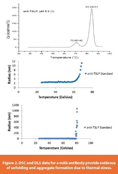
Figure 2 shows data from thermal stress testing of a mAb, anti-TSLP (Thymic Stromal Lymphopoietin) antibody, selected as a representative example of a mAb antibody.
DSC data (Figure 2 - top) show two peaks, the first at around 70°C with a second larger peak evident at around 84oC. These peaks indicate two discrete unfolding events, a plausible rationale being that one is associated with behavior in the Fab (antigen binding) region while the other is associated with the Fc region.3
DLS data (Figure 2 – middle) from a temperature ramp to around 70°C indicate that at temperatures above ~60°C the radius of the protein particles begins to increase, from around 2 to 10 nm. This is consistent with the formation of partially unfolded clusters. The second set of DLS data, from a higher temperature ramp (Figure 2 – bottom) shows that, in contrast, at around 80°C, there is a much more dramatic increase in particle size, to around 1000 nm (1 μm). This suggests substantially more significant aggregation. Conventional FTIR analysis was carried out to generate structural information to elucidate these observations (see Figure 3).
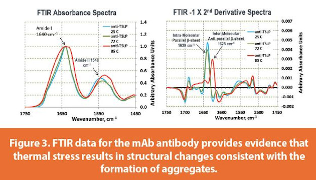
While FTIR data were gathered across both the Amide I and Amide II regions, changes in the Amide I wavelengths alone are sufficient to detect structural differences associated with the application of thermal stress. FTIR absorption spectra (Figure 3 - left) for samples stressed to 25°C and 72°C (blue and green traces respectively) are closely similar whereas the sample at 85°C generates a broader Amide I peak. Presenting the results in the form of -1 x 2nd derivative spectra (right), a standard strategy to resolve absorption peaks within a band, shows that this broadening is associated with the development of a peak at around 1625 cm-1; the wavelength associated with inter-molecular anti-parallel beta-sheet structure and by extension, the formation of aggregates.4,5,6,7,8 In contrast, the other two samples exhibit a peak at 1639cm-1 a wavelength correlated with intra-molecular parallel beta-sheet structure, an expected structural feature of the unaggregated mAb.
These data illustrate the ability of FTIR to detect the structural changes associated with aggregation. The absorbance of water in the Amide I region makes it difficult to get a good FTIR signal, necessitating measurement at relatively high concentrations. Here a concentration of 40 mg/mL was required which is far from representative of the few μg per mL typically associated with vaccine formulations.
Furthermore, FTIR measurements are manual rather than automated including sample loading and data processing; background and buffer spectra were measured separately for this system and then manually subtracted from the sample spectra. Overall measurement proved slow, difficult, manually intensive and temperature sensitive.
Detecting Aggregation with MMS
To compare the utility of MMS with conventional FTIR a follow-up study was carried out, investigating aggregation of a non-mAb protein antigen. In this study, control and heat stressed (65°C for 30 minutes) samples were measured at a concentration of 3 and 6 mg/mL. A control sample was also run at 10 mg/mL, creating 5 sets of data in total. Once the samples were loaded into the 24 well sample plate all aspects of analysis including data processing were automated. As a result, all the MMS measurements were completed in substantially less time than the single FTIR analysis reported in the preceding study, and with less manual input.
Absorbance spectra (Figure 4 left) show a broadening of the Amide I peak for the thermally stressed samples (orange and yellow traces) relative to the control samples. An interesting point to note is the very low levels of absorbance of the measurements. These samples were run at relatively low concentration and the absolute absorption was correspondingly low. However, signal to noise ratio was excellent and exceptional sensitivity was observed in the data, following automated buffer subtraction. The corresponding -1 x 2nd Derivative Absorbance plot provides better resolution of the Amide I peak. It shows that thermal stress results in a second peak at ~1620 cm-1 indicating the development of anti-parallel beta sheet structure and associated aggregation.
The results shown in Figure 5 illustrate the ways in which the MMS data can be automatically processed to enhance its informational value.
One approach is to use the Similarity or Area of Overlap tool (upper plot) which enables quantitative comparison of the 2nd Derivative spectra of a set of measurements, even if they are conducted at different concentrations. For this comparison the user assigns a baseline measurement, in this case the spectra measured for the 10 mg/mL control sample, and all other measurements are compared to it, following normalization of the area under the curve on the basis of concentration. Proteins with closely comparable structure will exhibit a high Area of Overlap, even when measured at different concentrations, while dissimilar proteins with be associated with lower values, even if measured at the same concentration.
Here the control samples are all closely similar with an Area of Overlap of 97.67 and 95.28% for the 6 mg/mL and 3 mg/mL samples, respectively. In contrast thermal stress reduces the Area of Overlap to 74.08 and 73.61% for the 6 mg/mL and 3 mg/mL samples. These results illustrate the simplicity and effectiveness of using Area of Overlap analyses to compare and quantify the level of aggregation in different samples.
Further insight into the structural changes induced by thermal stress can be gained from estimates of the quantities of different types of secondary structure in each sample. To generate these estimates the software automatically fits the 2nd derivative spectra for a specific sample to a collection of standard spectra with established structural forms. Figure 5 (bottom) shows the results which include estimates for the quantity of parallel beta sheet, unorganized, alpha helix, turn, and anti-parallel beta sheet in each sample. The level of anti-parallel beta sheet structure in the 3 mg/mL and 6 mg/mL samples increases to 29.5 and 28.2% respectively, relative to corresponding control values of 3.1 and 2.7%. These data directly quantify the extent of aggregation in terms of its impact on the secondary structure of the protein.
Conclusion
The unfolding and aggregation of antigens is detrimental to the safety and efficacy of vaccines and must be rigorously controlled to the point of delivery to the patient. The structural changes associated with aggregation can be detected from changes in IR adsorption across the Amide I band, with adsorption at ~ 1620 – 1625 cm-1 securely correlated with formation of the intermolecular anti-parallel beta-sheet structure observed in aggregates. Conventional transmission mode FTIR can detect this structure but only at high protein concentrations (~> 30 mg /mL). Equally importantly measurement is slow, complex and manual. In contrast, MMS can easily detect the formation of anti-parallel β-sheet formation at relatively low protein concentrations, in the range 1 to 6 mg /mL. Automated measurement, including advanced data processing, makes the technique well-suited to high-throughput screening and a valuable tool for vaccine development.
References
- Nabel, G.J. (2013) Designing Tomorrow’s Vaccines. N Engl J Med. 368(6), 551-560
- Wang, W., Roberts, C., J. (2013) Non-Arrhenius protein aggregation. AAPS J., 15, 840-851
- Ionescu, R.,M., Vlasak, J., Price, C., Kirchmeier, M. (2008) Contribution of variable domains to the stability of humanized IgG1 monoclonal antibodies. J Pharm Sci. 97, 1414-1426
- Sathya Devi, V., Coleman, D.R., Truntzer J., (2011) Thermal unfolding curves of high concentration bovine IgG measured by FTIR spectroscopy. Protein J, 30, 395-403
- Tian, F., Middaugh, C., R., Off erdahl, T., Munson, E., Sane, S., Rytting. J., H., (2007) Spectroscopic evaluation of the stabilization of humanized monoclonal antibodies in amino acid formulations. Int J Pharm., 335, 20-31
- Matheus, S., Friess, W., H.C. Mahler, H.,C., (2006) FTIR and DSC as analytical tools for highconcentration protein formulations. Pharm Res., 23,1350-1363
- van de Weert. M., Haris, P.I., Hennink, W.,E., Crommelin, D.,J., (2001) Fourier transform infrared spectrometric analysis of protein conformation: effect of sampling method and stress factors. Anal Biochem, 297, 160-169
- Dong ,A., Matsuura, J., Manning, M.,C., Carpenter, J.,F., (1998) Intermolecular beta-sheet results from trifl uoroethanol-induced nonnative alpha-helical structure in beta-sheet predominant proteins: infrared and circular dichroism spectroscopic study. Arch Biochem Biophys. 355, 275-281