If we want to scientifically understand the dynamics of LAL and rFC in terms of equivalency, then we must be able to sort out some conflicting data. Five broad claims have been made by a single LAL manufacturer against rFC which suggest that rFC underestimates endotoxin content. Here these five broad claims are refuted:
- Natural and non-microbiologically controlled water sources are an inappropriate matrix to demonstrate equivalence due to the LAL false positive Factor G pathway.
- Data that shows lower endotoxin results for rFC versus LAL invariably is derived from non-microbiologically controlled water including deionized water.
- LAL results do not always give the true answer in the presence of beta-glucans (βG), cellulosic residues or surfactant/zwittergent. Importantly, beta-glucan blocking buffers (βGBB) cannot completely negate these effects.
- Despite a recent LAL manufacturer’s claim that Factor B is needed in addition to Factor C to detect endotoxin, there is a simple proof that Factor C is the lone biosensor for endotoxin.
- The compendial validation requirements are contained in USP <1225> and require that such efforts are “fit for use” in that only drug products and substances going into injectable drug products are subject to endotoxin validation.
a). Natural and non-microbiologically controlled water sources are an inappropriate matrix to demonstrate equivalence due to the LAL false positive Factor G pathway.
The testing for endotoxin in pharmaceutical manufacturing begins with the testing of purified waters.1 Naturally sourced and non-microbiologically controlled water sources including prefiltration, carbon filtration or deionized water are not tested for endotoxin by pharmaceutical manufacturers. These waters remain largely uncharacterized in terms of microbiological content. Contents that affect a blood-based test like LAL includes various organic substances including glucans, cellulose, surfactants and detergents, all of which may skew test results. In many cases, such tests are in effect non-reproducible given that they are based upon a point in time that includes the uncontrolled environment from which they have come. Fungi which contain beta-glucans in particular are ubiquitous in the environment as responsible for the breakdown of organic materials in the ecosystem.
It is well documented that the potential contaminants sometimes found in pharmaceutical waters comes from biofilm2 present in purified water systems which is not the same thing as natural water contaminants that contain uncharacterized organic and microbiological substances in addition to endotoxin including beta-glucans and cellulosic residues. Most importantly, purified waters are monitored for total organic carbon (TOC) whereas potable waters including DI are not.
Sandle in “Characterizing the Microbiota of a Pharmaceutical Water System-A Metadata Study”3 conducted a fifteen year study of the microbiological quality of the three main types of waters listed as:
- Potable water sources (mains) > deionized – Bacteria are not the only microorganisms that inhabit source waters; there will be a complete ecosystem in operation which includes fungi, protozoans and algae.
- Purified water – product of reverse osmosis – biofilm and low level contaminants are Gram Negative bacteria.
- WFI - Water-for-Injection systems – almost no contaminants (endotoxin or bacteria)
Potable water is a natural feed water for purified and WFI water but is not controlled according to injectable drug standards (no endotoxin testing) and, importantly, is neither microbiologically or TOC controlled and contains beta-glucans as organic matter.
Sandle reviewed the quality of the various water classes (potable, purified, and WFI) in an extensive 15 year study. He described the water microbiological quality by water type. Bold added to highlight microbiological limits.
- Potable: Over the period of review, 1,040 samples were taken. Samples typically recovered microorganisms although few samples (201) were above the action level of 30,000 Colony Forming Units (CFU)/100 ml.
- Purified: The process of manufacturing the purified water was via reverse osmosis. For the study, 6,300 samples were tested. Of these, some 315 samples exceeded the action level of 100 CFU/100 ml (5%) and 347 isolates were recovered. The most common genera were “Pseudomonad type” organisms, with Ralstonia being the most prevalent.
- WFI: Few microorganisms are typically recovered from Water-for-Injection (WFI) systems. This is due to the nature of the method of producing the water (either reverse osmosis or distillation of purified water) and the distribution of the water, where the water is typically held at 80°C or higher..
- The review of data for the fifteen year time period shows that samples rarely exceed the specification for the water system (which is set by the pharmacopeia at 10 CFU/100 ml).
- Gram-negative bacteria are arguably the primary contaminants of WFI. From the 46,800 samples taken during the review period, only 300 samples detected Gram-negative rods (a rate of 0.6%) Of these 300 samples, 439 Gram-negative rods were recovered (less than two different organisms per sample.)
To aid the understanding of the microbiological quality of potable water Wenfa Ng4 gives a good visualization of the prevalence and variety of potable water contaminants, including DI water as shown in Figure 1.
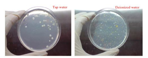
Figure 1. Microbes were recovered from deionized and tap water in significant numbers (with 0.1 g/L yeast extract) at 30°C after multiple days of incubation. From Ng, CC 3.0.
False activation of LAL via βG’s and cellulosic residues in portable water is inevitable and is not a phenomenon seen in purified and WFI waters. The additional cascade factor G activates the LAL clotting cascade via proclotting enzyme separate from endotoxin activation via Factor C.5
(b) Data that shows lower endotoxin results for rFC versus LAL invariably is derived from non-microbiologically controlled water including deionized water.
Data that has been used to demonstrate non-equivalence is from non-microbiologically controlled waters such as pre-filtration, carbon filter or deionized water. The often referenced Kikuchi study6 used both purified water containing naturally occurring endotoxins (NOEs) as well as naturally sourced waters (river, lake and sewer). This later set of waters has been repeatedly referenced by those desiring to show non-equivalence. But does it really show non-equivalence?
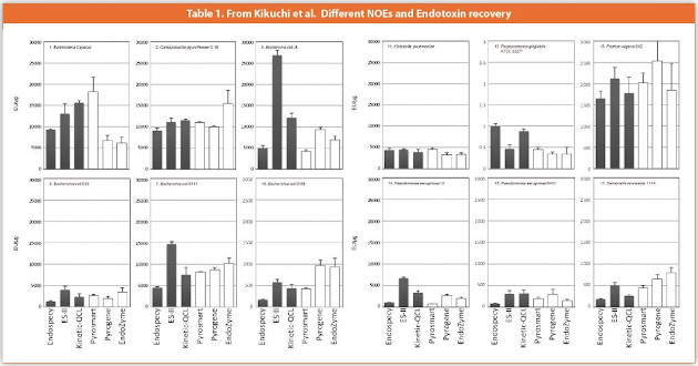
In terms of the recovery of specific bacterial types in purified water, the Kikuchi study showed similar recoveries for all organisms. There were some instances where LAL recovered more and some where rFC recovered more but results are overall comparable. Significantly, where one method set was better than the other the differences within the recoveries for the specific organism and methods showed significant divergence (LAL vs. LAL and rFC vs.rFC). These comparisons are shown in Table 1.
A couple of the organisms that appear to give better recovery using LAL are shown below: E. coli J5 and E. coli O111. Five of the six values for each data set agree except for the one over-recovery by one of the LALs using ES-II (turbidimetric). The issues that rFC cannot resolve is that one LAL doesn’t agree with another LAL or when LAL values are pushed up by the presence of βG’s or cellulosic residues.
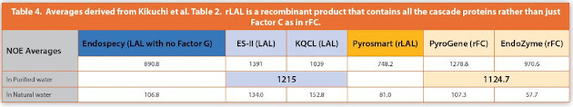
The next Kikuchi table (Table 2) below (Table 3 here) used NOE as added to purified water (rows 1-5) and also various “natural waters” including those from rivers, lakes, and sewage (rows 6-11). The consistency and equivalence of the data can be seen intuitively without statistical analysis for both sets. The simple averages are provided in Table 4.
From derived Table 4 below we can see that LAL and rFC determinations for NOE in purified water agree while the values obtained when testing endotoxin in natural waters tend to show higher endotoxin values with LAL relative to rFC. This can be attributed to the presence of βG’s, cellulose, and/or surfactant/detergents in natural waters. According to Roslansky and Novitsky7 the presence of any of these can contribute to an increase in sensitivity of LAL response due to the Factor G pathway. Blood systems, even of primitive animals, are extremely complex matrices and likely contain a dozen proteins (8 of which are known to be part of the LAL cascade).a The contention that the use of βGBB’s can block all non-endotoxin reactivity in the LAL cascade will be examined in the next section.
The Endospecy product which does not contain Factor G shows significantly lower results than either the ES-II or KQCL LAL values indicating that these samples likely do contain β-glucans which makes the ES-II and KQCL values likely inflated. In general, the NOEs in purified waters produce very close recoveries (LAL vs. rFC) whereas NOEs in natural waters typically show more activity with LAL for the reasons discussed here. Bolden and Smith also demonstrated the equivalence of rFC and LAL using purified water relevant bacteria in actual pharmaceutical buffer systems.8
c). LAL results do not always give the true answer in the presence of beta-glucans (βG), cellulosic residues or surfactant/zwittergent. Importantly, beta-glucan blocking buffers (βGBB) cannot completely negate these effects.
Lost in the sudden deluge of comparability data intending to compare LAL to rFC are the inherent differences seen when comparing various LAL results. Similarly to the results shown in Table 2 above, LAL often can be seen to differ LAL to LAL, and often to a degree equal to or larger than the difference seen between LAL and rFC test comparisons. Invariably, the data used to claim non-equivalence comes from non-microbiologically controlled water sources and, in some cases, uses a limited set of LAL types that are expected to better agree with each other.

More research is needed on βGBB efficacy in overcoming LAL false positive results and the widespread use of a “glucan blocking buffer”. It is mostly assumed by industry participants that βGBB will provide 100% knock out of the effects of βG’s when using LAL. However, the study by Roslansky and Novitsky showed that the method of extraction of lysate during LAL manufacture either by using chloroform extraction, addition of zwittergent (surfactant) or both, has a significant effect on both endotoxin and glucan sensitivity and causes LAL results to differ significantly from one another.
Furthermore, the addition of cellulosic residues from naturally sourced waters adds an activity that is different from that of conventional 1,3-D-β-glucan. This was seen in the Roslansky and Novitsky study where the test recovery is seen to differ depending upon the antiglucan enzyme treatment used glucanase (1,3 beta glucans) or cellulase (1,4 beta glucans). Cellulosic residues can be expected to occur in natural source waters as the breakdown products of fungi, grass, wood, and various other plant derived materials. The issue of cellulosic breakdown products was first seen in pharmaceutical manufacturing samples and called LAL reactive material (LRM). The blocking of 1,3 beta glucans is not the same as the blocking of 1,4 beta glucans as represented by cellulose in the Roslansky and Novitsky study.

Figure 2. Cellulose is a (1-4) β -D-glucan composed of repeat glucose units (left). β-glucans are (1-3) β-D-glucans connected at carbon atoms 1 and 4 (right). Cellulase breaks the cellulosic bonds while laminarinase/glucanase breaks (1-3) β -glucan bonds rendering them LAL inactive. Alternatively, βGBB seeks to displace 1-3 β-D-glucans by occupying the Factor G receptors in LAL. There are a myriad variety of forms of glucans (branching etc.) and one can expect some natural forms to have higher affinity to Factor G than the common forms used in blocking buffers.
Some maintain that only 1-3 β-D glucans are LAL reactive and that cellulosic residues are not. However, Henne et al. showed that “aqueous extracts of cellulose hollow fibers (CHF) exhibit positive reactions in some Limulus amebocyte lysate (LAL) tests”, where “oxidative or acidic degradation of cellulose does not result in the formation of LAL-reactive material (LAL-RM). On the other hand, sterile cotton wool shows LAL reactivity, and cellulose acetate regains LAL reactivity when it is saponified. Thus, it appears likely that the LAL-RM found in CHF is of purely cellulosic origin and cross reacts with a number of commercially available lysates.”9 Nagasawa et al. draw a similar conclusion: “we cannot predict the biological activity of (1-3) β-D-glucan from cellulose materials, because its structural features are not clear.”
The Roslansky and Novitsky study presents compelling evidence that one LAL does not always equal another LAL. This can be seen in reactivity to both various Glucans as well as Zwittergent content as commonly added to LAL in the LAL manufacturing process.
In Table 5, the 0.02% and 0.03% zwittergent conc. shows that the difference in LAL recovery is 1.56 versus 6.25 pg/ml (a four-fold difference). At 0.04% zwittergent content, the endotoxin recovery for the same LAL test has swelled to 25 pg/mL a 16X differential versus that containing 0.03% zwittergent.
Roslansky and Novitsky describe the LAL activity of cellulose residues as a separate phenomenon from that of (1-3) β-D Glucans:
By using the gel-clot method and Pyrotell, the activity of LAL-RM was reduced from 10 to 0.125 ng/ml after digestion with cellulase. The amount of activity left could be attributed to the intrinsic endotoxin contamination of the cellulase, since water plus enzyme had activity nearly identical to that of the digested LAL-RM. Cellulase had no effect on endotoxin. This enzyme reduced the activity of laminarin from 8,000 to 2,000 ng/ml and, therefore, is most likely contaminated with a trace amount of a P-(1,3)-hydrolase. The digestion experiments support the contention that LAL-RM is a glucan with primarily β-(1,4) linkages and that it is different from laminarin, which has predominantly β-(1,3) linkages.
LAL reactive material was also found to be associated with hemodialysis and dialyzer filtration and identified as a cellulosederived mixture.11,12
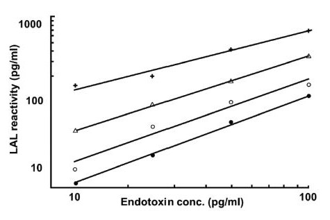
Figure 3. Enhancement of standard endotoxin lines by three concentrations of curdlan: 250 ng/mL (+), 25 ng/ml (∆), and 2.5 ng/ml (○). No curdlan was added to the standard line (●). Endotoxin standards were spiked with curdlan and were assayed by the kinetic turbidimetric method. Derived from Roslansky & Novitsky.
A simple set of data generatedb using today’s available LALs supports the findings of Roslansky and Novitsky as shown in Table 6. A natural local water was tested by rFC and three different LALs with and without βGBB. Enzymatic treatment of the water prior to LAL testing was also performed in lieu of βGBB, as per the method described by Roslansky and Novitsky. This included treatment with either glucanase or cellulase. The results show the exaggeration of results using LAL when testing natural water due to glucan and/or cellulose effects. The use of βGBB for natural (source) waters only reduces a fraction of the non-endotoxin effect. The dynamics shown here rarely if ever come into play in pharmaceutical manufacturing because non-microbiologically controlled waters are never tested except as requested by LAL suppliers for rFC – LAL equivalence studies.
A second simple experiment shows, via a different method, that natural waters are not a suitable matrix for comparison studies. LAL gave an initial result of 28.3 EU/mL. After addition of 100 EU/mL of RSE the test was performed again and the result was 464 EU/mL. Use of βGBB reduced the result slightly to 326 EU/mL, but glucanase treatment of this sample prior to addition of the 100 EU/mL RSE gave 133 EU/mL.
However, when rFC was tested an initial value of 18 EU/mL was obtained. When 100 EU/mL of RSE was added to this water and the test was repeated, the result was 132 EU/mL.
Subscribe to our e-Newsletters
Stay up to date with the latest news, articles, and events. Plus, get special offers
from American Pharmaceutical Review – all delivered right to your inbox! Sign up now!
The synergistic effects of natural water containing glucans on endotoxin are obvious. Eff orts to confound equivalency tests and present the data as “pharmaceutically relevant” should be viewed as purely commercial. The variance seen is not because the endotoxin is “natural” or “purifi ed” endotoxin as here both have acted similarly. This is not a matter of rFC “under detecting” endotoxin, rather it is the case of LAL exaggerating the results due to beta-glucan-endotoxin synergistic eff ects, an eff ect which does not occur in the testing of purified waters.
d) Despite a recent LAL manufacturer’s claim that Factor B is needed in addition to Factor C to detect endotoxin, there is a simple proof that Factor C is the lone biosensor for endotoxin.

Figure 4. Large granules within the amebocytes contain all the Limulus clotting cascade factors(left). To release the factors, Factor C exists on the granulocyte surface. When Factor C contacts LPS then the contents of the granules are released into the hemolymph (called exocytosis). At right, the empty amebocytes after exocytosis (degranulation) are shown(right). From Armstrong.13
Most recently one LAL manufacturer has made the claim that not only Factor C but also Factor B is needed for detecting endotoxin. The longstanding view is that Factor C is the lone natural “biosensor” for endotoxin. There are more than a few contradictions in this new view (that you need Factor B to detect endotoxin) but there is a simple explanation as to why it is false.
According to Wang, Ho and Ding: “Circulating Factor C derived from hepatocytes binds Gram-negative bacteria or LPS and triggers a further exocytosis of cellular Factor C from the large granules of amebocytes. The LPS activated Factor C initiates the serine protease cascade.”14 This can also be seen in the Koshiba et al. fi gure below. See also Cerenius and Söderhäll15 and as described here by Ariki et al.16

The exocytosis of clotting system components is initiated by the binding of LPS to the membrane-associated form of factor C. Factor C is an LPS-responsive serine proteinase zymogen. It is present, as are the other components of the cascade, in the large granules of hemocytes. In addition, this protein is present in discrete areas in the cell membrane. Binding in membrane-associated factor C results in the exocytosis of coagulation system components including more factor C. This activation has been likened to the activation of platelets by thrombin through proteinase-activated receptors.
The Koshiba et al. figure below shows that it is the interaction between Lipid A and the “tripeptide motif” of Factor C that utilizes the precise spacing of tryptophan and lysine hydrophobic amino acids that is responsible for endotoxin detection. Significantly, Factor B is not released until Factor C exocytosis brings about the release of LAL cascade factors from the large granules in Limulus blood. Therefore, it is Factor C that is the biosensor that determines if endotoxin is present in the hemolymph milieu before Factor B is even released into the milieu. If Factor B were needed to bind endotoxin, then the release of cascade factors could not occur.
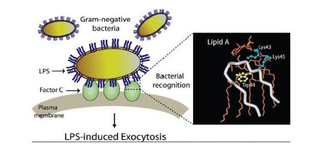
Figure 5. from Koshiba et al.17 Model of bacterial recognition by Factor C. The recognition of LPS on Gram-negative bacteria by membrane-bound Factor C initiates the horseshoe crab innate immune response by releasing all cascade factors into the hemolymph.
The old adage that “correlation is not causation” applies here. If Factor B touches Factor C during LPS binding (after it has been let out of the granule by Factor C) that does not suggest that rFC numbers should be lower than LAL numbers (the purpose for proposing the idea in the first place). In fact it suggests the opposite, namely as Masakazu Tsuchiya stated in his article18 it would make LAL more selective than rFC and thus would bind fewer not more LPS molecule types: “These findings suggest that the role of Factor B in the cascade system is not only to activate proclotting enzyme, but to increase specificity of LAL to endotoxin.”
e). Compendial validation requirements are contained in USP <1225> and require that such efforts are “fit for use” in that only drug products and substances going into drug products are subject to validation.
Validation efforts should be “fit for use” or “fit for its intended purpose”19 in that the validation pertains to that which is to be tested routinely. Non-microbiologically controlled waters are not a part of routine endotoxin testing. Significant efforts have gone into validating the various commercial rFC products now available including supplier validations as well as numerous drug manufacture performed validations, some of which have resulted in FDA approval for marketed end-products. These validation efforts do not employ river, lake, sewer, pre-filtration, carbon bed, or deionized water which are not within scope for endotoxin testing in pharmaceutical manufacturing. Science, by definition, must be reproducible, otherwise, it is dependent upon the single anecdotal instance from which it was generated. This is to say that the content of nonmicrobiologically controlled water will vary greatly. Scientifically, validation test materials must be highly characterized and provide a reproducible matrix for confirmatory studies as well as support the actual articles to be placed under routine test.
According to USP General Notices 3.10.10:
- Applicability of Standards to Drug Products, Drug Substances, and Excipients
- The applicable USP or NF standard applies to any article marketed in the United States that (1) is recognized in the compendium and (2) is intended or labeled for use as a drug or as an ingredient in a drug.
Natural and other non-microbiologically controlled waters do not fit into a relevant category for compendial testing. Endotoxin test users are free to follow existing compendial requirements for validation of relevant articles including purified waters as stated in USP <1225> to validate their products for routine test or may follow EP 2.6.32 which, like USP <85>, requires only product suitability testing (inhibition / enhancement testing). The addition of a new USP informational-only endotoxin chapter will not alter USP <1225> requirements.
Conclusion
These studies taken together make it clear that the testing of nonmicrobiologically controlled waters using various LALs cannot produce a “gold standard” result, even when using βGBB, due to the background presence of beta-glucans. Purified waters which are monitored for TOC do not have this organic backdrop. The temptation to include data from source waters in an effort to compare “natural endotoxin” should be resisted or should at least include enzymatic treatment of such waters given that βGBB provides only a partial reduction of non-endotoxin effects on LAL. The testing of purified waters provides the appropriate test matrix to achieve “gold standard” results when endotoxin test comparisons are desired.
Author Biography
Kevin Williams spent 30 years at Eli Lilly & Company developing endotoxin assays and detection technology in the QC lab. He then worked at Hospira(now Pfizer), Lonza, GE Water before moving to bioMérieux. He has authored several books on endotoxin (Endotoxins second and third edition, Informa) including, most recently Endotoxin Detection and Control in Pharma, Limulus, and Mammalian Systems (2019, Springer Nature).
References
- Water for Pharmaceutical Use, FDA, current as of 08/27/2014, accessed 07/09/2020.
- Tim Sandle, The Problem of Biofilms and Pharmaceutical Water Systems, Amer. Pharm. Rev., Friday, December 22, 2017, accessed July 9, 2020.
- Sandle T (2015) Characterizing the Microbiota of a Pharmaceutical Water System-A Metadata Study. SOJ Microbiol. Infect Dis 3(2): 1-8.
- Ng W. 2018. Microbes in deionized and tap water: Implications for maintenance of laboratory water production system. PeerJ Preprints 6:e181v6 https://doi.org/10.7287/peerj.preprints.181v6.
- Hodes, D. S., D. Heon, A. Hass, A. C. Hyatt, and H. L. Hodes. 1987. Reaction of fungal products with amebocyte lysates of the Japanese horseshoe crab, Tachypleus tridentatus. J. Clin. Microbiol. 25:1701-1704.
- Kikuchi et al., Collaborative study on the bacterial endotoxins test using recombinant Factor C-based procedure for the detection of lipopolysaccharides, Pharm. And Med. Device Regulatory Science, Vol. 48, No. 4, 252-260 (2017).
- Roslansky and Novitsky, Sensitivity of Limulus amebocyte lysate (LAL) to LAL-reactive glucans, Associates of Cape Cod, Inc., Box 224, Woods Hole, Massachusetts 02543, JOURNAL OF CLINICAL MICROBIOLOGY, Nov. 1991, p. 2477-2483.
- Application of recombinant Factor C reagent for the detection of bacterial endotoxins in pharmaceutical products, Bolden and Smith, PDA Journal of Pharmaceutical Science and Technology September 2017, 71 (5) 405-412.
- Henne et al., Hollow-fiber dialyzers and their pyrogenicity testing by Limulus amebocyte lysate, Artificial Organs, Aug;8(3):299-305, 1984.
- Nagasawa et al., Experimental proof of contamination of blood components by (1-3)-β-D-glucan caused by filtration with cellulose filters in the manufacturing process, Jour. Artificial Organs (2003) 6:49–54.
- Pearson FC, BohenJ, Lee W, Bruszer G, Sagona M, Dawe R, Jakubowski G, Morrison D, Dinarello C. Comparison of chemical analyses of hollow-fiber dialyzer extracts. Artificial Organs 1984; 8: 291- 298.
- A Study on Limulus Amebocyte Lysate (LAL) Reactive Material Derived from Dialyzers,YOSHIOKA et al., Japanese Jour. Surgery, VOL 19, No. 1 pp. 38-41, 1989.
- Peter B. Armstrong, MOTILITY OF THE LIMULUS BLOOD CELL, J. Cell Sci. 37, 169-180 (1979).
- Wang, Ho and Ding, Transcriptional Regulation of Limulus Factor C, THE JOURNAL OF BIOLOGICAL CHEMISTRY, Vol. 278, No. 49, Issue of December 5, pp. 49428–49437, 2003.
- Lage Cerenius Kenneth Söderhäll, Coagulation in Invertebrates, Journ. Innate Immunity, Published online: November 6, 2010, accessed 7/11/2020.
- Ariki et al., A serine protease zymogen functions as a pattern-recognition receptor for lipopolysaccharides, Proc Natl Acad Sci., 2004, Jan 27;101(4):953-8.
- Takumi Koshiba, Tomoyuki Hashii, and Shun-ichiro Kawabata, A Structural Perspective on the Interaction between Lipopolysaccharide and Factor C, a Receptor Involved in Recognition of Gram-negative Bacteria, . THE JOURNAL OF BIOLOGICAL CHEMISTRY VOL. 282, NO. 6, pp. 3962–3967, February 9, 2007.
- Masakazu Tsuchiya, INNOVATIVE MECHANISM OF LIMULUS AMEBOCYTE LYSATE ACTIVATION TO ACHIEVE SPECIFICITY AND SENSITIVITY TO ENDOTOXIN; COMPARISON WITH RECOMBINANT FACTOR C REAGENTS, Microbial Solutions, Charles River, Charleston, SC, USA, International Journal of Development Research Vol. 10, Issue, 06, pp. 36751-36756, June, 2020.
- Analytical Procedures and Methods Validation for Drugs and Biologics Guidance for Industry, Guidance for Industry, FDA (HHS/CDER/CBER) 2015.
a Including the following proteins: Factor C, Factor G, Factor B, proclotting enzyme, coagulogen, and the three serine protease inhibitors LICI 1, 2, and 3.
b Preliminary data being followed up on in a larger study