What are Endotoxins?
Endotoxins are lipopolysaccharides (LPS) embedded in the outer membrane of Gram-negative bacteria. They represent pyrogens that are of the greatest consequence to manufacturers of parenteral solutions and medical devices. Endotoxins are consequential because they are environmentally ubiquitous, heat stable, and filterable. Importantly, they are extremely pyrogenic. Endotoxins stimulate monocytes and macrophages to secrete an array of inflammatory cytokines that include tumor necrosis factor alpha (TNFα), the interleukin (IL)-1 family, IL-6, IL-8, the IL-10 family, the IL-12 family, the IL-15 family and the transforming growth factor beta (TGFβ).1
Normally, there are three (3) structural components to LPS. Lipid A, the hydrophobic portion of LPS, a hydrophilic heteropolysaccharide core region, and the O-specific oligosaccharide region that differs from strain to strain within a serotype. The first sugar of the core region, (3-deoxy-D-manno-octulosonic acid, or KDO) is attached to the 6’ position and connects the core polysaccharide to Lipid A. All of the biological activity of LPS resides in the Lipid A region.
Lipid A has a beta diglucosamine backbone with phosphoryl groups at positions 1 and 4’. For E. coli and other members of the Enterobacteriacea family of Gram-negative bacteria, this backbone is acylated with beta-hydroxymyristate at the 2, 3, 2’, and 3’ positions. Secondary acyl groups are esterified with laurate and myrisitate at the 2’ and 3’ locations respectively. The Lipid A architecture, described here, is the classical structure and is one of the most powerful activators of the innate immune response.2
Not all Gram-negative organisms produce the Lipid A structure that activates the innate immune response. Activation depends on the presence of phosphoryl groups, the number acyl chains, and the length of the acyl chains. LPS from Helicobacter pylori, Yersinia pestis, and Francisella tularensis are pathogenic, yet their LPS structures are poorly recognized by Toll-like receptors (TLR4) and thus evade host detection.3,4 Even among the Enterobacteriacea family, the classical LPS architecture is subject to change. Palmitate additions and modifications of the Lipid A phosphate groups with cation sugars or phosphoethanolamine occur under conditions that are low in magnesium and pH. Modifications also occur in high iron, aluminum, and calcium environments. LPS from all Gram-negative bacteria undergo constant modifications or remodeling. The remodeling of Lipid A is determined by environmental conditions and is largely controlled via the PhoP-PhoQ and PmrA-PmrB two-component regulatory systems.5
The outer membrane of Gram-negative bacteria is highly asymmetrical, containing glycerophospholipids in the inner leaflet and LPS that is exposed to the cell surface. It serves as a permeability barrier. Most nutrients pass through this barrier via a family of outer membrane proteins (OMP) called porins. Alterations to the outer membrane affects its integrity, fluidity, and permeability.6 The remodeling of LPS and the outer membrane components represent a coordinated means of survival. Outer membrane vesicles (OMV) have a Gram-negative bacterial secretory process that facilitates remodeling of the outer membrane.7

Outer membrane vesicles are nanoparticles that are universally produced by Gram-negative bacteria. They range from 20 – 250 nm in diameter and contain an array of proteins, lipids, LPS, and other biologically active substances that are no longer needed or that are detrimental to survival. They typically bear a composition that is similar to that of the outer membrane of the bacteria that elaborated the OMV. OMV production is a bacterial stress response and has an important role for nutrient acquisition, biofilm development, horizontal genetic exchange, and other interspecies interactions that occur in complex microbial communities.8 These communities are often under temperature and nutritional stress. Endotoxins from these microbial communities cannot be created in a laboratory as there are seasonal and other indigenous factors that impart local distinctiveness. These microbiological populations exist in all parenteral pharmaceutical operations.
The Past
In 1912, Hort and Penfold developed and refined a rabbit animal model that advanced the study of injection fevers. Their rabbit model meticulously controlled the breed, sex, weight, food, and environmental husbandry conditions. In every experiment, dosing was reported. Thermometric measurements were taken 30 minutes apart. The experimental design allowed them to conclusively demonstrate that fever-producing bodies (FPB) were absent when freshly distilled water was used for intravenous injections. Hort and Penfold dispelled all of the theories that were associated with: (a) water-fever, (b) salt-fever, (c) carbohydrate-fever, (d) ferment-fever, and (e) tissue-fever.9 Unfortunately, it wasn’t until 1923 that their work was appropriately recognized.
In 1923, Florence Seibert acknowledged the fundamental value of the scientific work by Hort and Penfold.10 Her investigations of injection fevers used the same rabbit model they developed. Seibert successfully confirmed and then extended the premise that pyrogenic materials in distilled water are of bacterial origin and that these materials are not removed by filtration. Most importantly, she presented “a method for the preparation of a non-pyrogenic water” (her words). Her method of preparing non-pyrogenic distilled water involved the heating of connection stoppers several times with alkali (depyrogenation) and introducing a “trap” above the distillation flask. The trap prevented the pyrogens from being mechanically carried over into the distillate. Her invention – now called baffles or demisters – removed entrained pyrogen-containing water droplets that are found on all modern vapor compression and multi-effect distillation equipment.
With the emergence of an entire parenteral industry in the 1930s and the large demand for parenteral solutions prior to and during World War II, the need to assure that commercial parenteral solutions were free from pyrogen contamination triggered the United States Pharmacopeia (USP) to develop a compendial test for pyrogens. In 1941, the USP, NIH, FDA, and 14 pharmaceutical companies collaborated in the very first study on pyrogen testing. The study utilized the rabbit model that was developed by Hort, Penfold, and Siebert and the results from the collaborative effort led to the inclusion of the first compendial pyrogen test in the 12th edition of the USP in 1942. The test required intravenous injection of test solution and the measurement of the rectal temperature of a group of rabbits for three hours. The USP <151> Rabbit Pyrogen Test (RPT) was the sole method of assessing the pyrogenicity of intravenous fluids and relevant medical devices for over 40 years.
During the period of 1964 to 1968, Levin and Bang reported that Gram-negative bacterial endotoxins have the capacity to coagulate the blood of the Atlantic horseshoe crab, Limulus polyphemus. They determined that the horseshoe crab’s blood sensitivity to bacterial endotoxins – derived from Gram-negative bacteria – was due to enzymes that are within their blood cells, amebocytes.11 Since the test was based on the blood of the horseshoe crab (Limulus) and since the enzymes were obtained from the Limulus amebocytes by lysis with high purity distilled water, the test was named Limulus Amebocyte Lysate, or LAL. A 1971 publication by Levin and Cooper reported that LAL was more sensitive and less costly than the RPT.12 It prompted worldwide interest in determining the suitability of the BET as a replacement for RPT.
Subscribe to our e-Newsletters
Stay up to date with the latest news, articles, and events. Plus, get special offers
from American Pharmaceutical Review – all delivered right to your inbox! Sign up now!
In 1973, the FDA published proposed manufacturing specifications for the production of LAL. Manufacturers of LAL were required to submit samples of each batch for testing by the FDA prior to its use. Provisional use of LAL allowed the industry to obtain real-world experience. The innovative LAL test was routinely used to test raw materials, in-process samples, First of Batch (FOB), and Middle of Batch (MOB) samples. End of Batch (EOB) samples required rabbit testing as the EOB was considered the worst case.
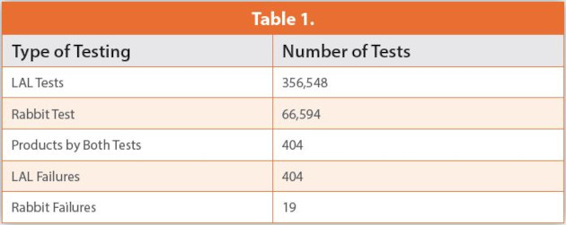
Recognizing the simplicity and robustness of the LAL test, Baxter Travenol, the largest producer of medical devices and LVPs, made a corporate decision in 1973 to validate LAL as an endotoxin test for their global facilities. In 1979 and 1982, they reported their annual endotoxin testing results.13,14 These data included 66,594 USP rabbit pyrogen tests and 356,548 LAL tests. Notably, 404 naturally contaminated samples were tested by both methods. The LAL test confirmed the presence of endotoxin, yielding a test failure for all 404 samples. However, following USP Chapter <151> Pyrogen Test, eight rabbits tests with all 404 samples resulted in only 19 failures. These data unequivocally demonstrated that LAL was the more sensitive method and that Gram-negative bacterial endotoxins were the pyrogen of greatest consequence to parenteral product manufacturing. These data and 45 years of experience attest to the specificity (the ability to detect a range of endotoxins) of the LAL test. As represented in Table 1; a combined summary from Table 6 Mascoli and Weary (14) and Table 7 Pearson, Weary, and Dabbah (13).
The Present
Commercial recombinant proteins are being advanced as a replacement to naturally sourced LAL reagents currently defined in the harmonized pharmacopeia. Three products are based on Factor C, the first endotoxin binding factor in the LAL cascade. Recombinant products are also being developed that contain all three of the clotting factors from the Asian (Tachypleus) and American (Limulus) horseshoe crab species. Studies purporting to demonstrate equivalency between the current compendial methods and the recombinant methods have been largely predicated on a comparative detection of purified LPS15,16 or on clean samples of Water for Injection (WFI).17 These studies, however, do not address the issue of specificity, i.e., the capability of detecting a range of natural environmental endotoxins.
Several studies have compared rFC to that of LAL using environmental endotoxins. Thorne et al., examined air samples from 10 livestock production facilities.18 The study design, sample size, and statistical evaluations were remarkable. However, air samples from livestock facilities do not represent pharmaceutical manufacturing environments. Additionally, the endotoxins associated with the bacteria in this sampling were more likely representative of the intestinal tract, not the non-fermenters that typically inhabit water systems. Kikuchi did examine environmental endotoxins from a variety of water sources.19 The sample set was very limited but despite that, the data indicated that recombinant methods can underpredict endotoxin concentrations with naturally contaminated waters.
Akers et al. recently compared all current publications purporting to compare rFC to LAL.20 Their review concluded that all data currently available demonstrates suitability of rFC but does not demonstrate comparability. They note most of the published studies claiming comparability include data from test articles that have no measurable autochthonous endotoxin activity in any segment of the manufacturing process. It is not possible to claim comparability when the impurity that is being measured, in this case, endotoxin activity, is absent in the test article at quantifiable levels. The recovery of the analyte (RSE or CSE) does not experimentally confirm the alternative method’s ability to recover natural product contaminants.
Charles River recently completed an evaluation that simultaneously examined three commercially available recombinant Factor C products, a single recombinant LAL cascade that is under development, and two FDA licensed LAL reagents. Pharmaceutical pre-treatment water samples, from parenteral manufacturing facilities in Europe, were used to assess the specificity of recombinant reagents. This study (due to be published) utilized a statistically relevant sample set. It demonstrated a propensity of all recombinant reagents to underestimate natural endotoxin concentrations. The underestimation was not due to a glucan bias of the LAL reagents since carboxymethylated curdlan was used as a glucan blocker for Charles River LAL reagent and both LAL reagents yielded similar results. Additionally, glucan-specific assays demonstrated that the glucan concentration of 95% of the samples was less than 10 pg/mL.21 The results of this study demonstrated that all recombinant products require additional development in order to detect the fever-producing bodies (endotoxins) identified by Hort, Penfold, and Seibert over 108 years ago.
The Future
The ability to measure and detect all of the endotoxins that are produced by the microbial community that inhabit purified water systems (members of Burkholderiaceae, Methylobacteriaceae, Comamonadaceae class / family) is absolutely essential for operational control of a WFI system. At the present time, recombinant alternatives to the LAL test are not consistently capable of doing so. Further advances in recombinant protein chemistry and/or recombinant formulations must be made. Refinements to recombinant alternatives to LAL must consider reactivity to natural environmental endotoxins instead of highly purified lipopolysaccharide standards. Discrepancies between the compendial LAL test and any recombinant alternatives must be scientifically resolved. Similar to the adoption of the LAL test in the early 1980s, comparative pyrogen testing must be done to address the ultimate concern – patient safety.
LAL is not a single enzyme. LAL is not three enzymes. LAL does contain three protease zymogens that are known to be involved in the sequential activation by bacterial endotoxins. LAL also contains the Factor G enzyme that is sensitive to beta 1-3 glucans. LAL contains the soluble protein coagulogen which is converted to in insoluble gel by the activated Proclotting Enzyme. LAL also contains three serpin like proteins that regulate the LAL endotoxin and glucan activation cascade. LAL contains an Anti-LPS Factor that neutralizes bacterial endotoxins, anti-microbial peptides, alpha2-macroglobulin, big defensins, and cystatin. LAL is a primitive immunological system that has served the horseshoe crab for 450 million years, and humans for the last 40. Limulus polyphemus will continue to serve as an efficient and reliable safety test for human and animal injectable drugs. We recognize that there are concerns pertaining to the sustainability of the Atlantic horseshoe crab population. Charles River has developed a cartridge based microfluidic LAL test that refines and reduces the Limulus resource requirement by 95%. Charles River, and all of the FDA licensed manufacturers of LAL, have for decades instituted conservation and educational programs that will ensure the preservation of this remarkable species.
Author Biography
John Dubczak is the Executive Director for Reagent Development for the Microbial Solutions division of Charles River Laboratories. For the last 20 years, he was the General Manager and responsible for the production and technical operations for the Endosafe® brand in Charleston, SC. Prior to joining Charles River, John was a long-term employee of Baxter Healthcare Corp., where he developed Baxter’s proprietary LAL formulation and manufacturing process. As a member of the R&D team, he also developed methods for product testing and explored the clinical applications of LAL. With seven years of Large Volume Parenteral manufacturing experience, he brings an in-depth understanding of issues surrounding all aspects of endotoxin and microbiological testing.
References
- Li, Wei., et al. Lipopolysaccharide-Induced Profiles of Cytokine, Chemokine, and Growth Factors Produced by Human Decidual Cells Are Altered by Lactobacillus rhamnosus GR-1 Supernatant. Reproductive Sciences 2014, Vol.2(7) 939-947.
- Caroff, M., Karibian, D. Structure of Bacterial Lipopolysaccharides. Carbohydrate Research 338 (2003) 2431-2447.
- Golenbock, D.T., Hampton, R.Y., Qureshi, N., Takayam, K., Raetz, C.R., Lipid A-Like molecules antagonize the effects of endotoxins on human monocytes. J Biol Chem 1991; 266: 19490 -19498.
- Ancuta, P., Pedron, T., Sandstrom, G., Chaby, R., Inability of the Francisella tularensis lipopolysaccharide to mimic or to antagonize the induction of cell activation by endotoxins. Infect Immun 1996; 64: 2041 – 2046.
- Chen, D.H., Groisman, E.A., The Biology of the PmrA/Pmrb Two Component System: The Major Regulator of Lipopolysaccharide Modifications. Annu Rev. Microbiol, 2013; 67: 83-112.
- Kulp, A., Kuehn, M., Biological Functions and Biogenesis for Secreted Bacterial Outer Membrane Vesicles. Annu Rev. Microbiol, 2010; 64; 163 – 184.
- Bonnington, K.E., Kuehn, M.J., Outer Membrane Vesicle Production Facilitates LPS Remodeling and Outer Membrane Mainatenance in Salmonella during Environmental Transitions. mBio September / October 2016 Volume 7; Issue 5; e01532-16.
- Schwechheimer, C, Kuehn, M.J., Outer-membrane vesicles from Gram-negative bacteria: biogenesis and functions. Nat Rev Microbiol, 2015; 13(10): 605-619.
- Hort, E,. Penfold, W.J., A Critical Study of Experimental Fever. Lister Institute of Preventive Medicine, March 14, 1912.
- Siebert, F. Fever Producing Substance Found in Some Distilled Waters. Sheffield Laboratory of Physiological Chemistry in Yale University, New Haven, Conn. September 8, 1023.
- Levin, J.; Bang, F. B. The Role of Endotoxin in the Extracellular Coagulation of Limulus Blood. Bull. Johns Hopkins Hosp. (1964) 115:265–274.
- Cooper, J. F.; Levin, J.; Wagner, H. N. New, Rapid, in-Vitro Test for Pyrogen in Short-Lived Radiopharmaceuticals. Journal of Nuclear Medicine; Society of Nuclear Medicine, 1970; Vol. 11, pp 273–287.
- Pearson, F.C., Weary, M.E., and Dabbah, R. “A Corporate Approach to In-Process and End-Product Testing With the LAL Assay for Endotoxin,” Endotoxins and Their Detection With theLimulus Amebocyte Lysate Test, 1982, Alan Liss, 150 Fifth Avenue, New York, New York,10011.
- Mascoli, C.C.,and Weary, M,E. “Applications and Advantages of the Limulus Amebocyte Lysate (LAL) Pyrogen Test for Parenteral Injectable Products”. Biomedical Applications of the Horseshoed Crab (Limulidae), 1979, Alan Liss, 150 Fifth Avenue, New York, New York.
- Abate, W., et al. “Evaluation of Recombinant Factor C Assay for the Detection of Divergent Lipopolysaccharide Structural Species and Comparison with Limulus Amebocyte Lysate- Based Assays and a Human Monocyte Activity Assay.” Journal of Medical Microbiology 66 (2017): 888–897.
- Muroi, M., Ogura, N., Mizumura, H., Aketagawa, J., Oda, T., Tanamoto, K., Application of a Recombinant Three-Factor Chromogenic Reagent, PyroSmart, for Bacterial Endotoxins Test Filed in the Pharmacopeias. Bio. Pharm. Bull. 42, 2024-2037 (92019).
- Bolden, Jay. 2020. Application of Recombinant Factor C Reagent for the Detection of Bacterial Endotoxins in Pharmaceutical Products and Comparability to Limulus Amebocyte Lysate. Pharmacopeial Forum, Stimuli to the Revision Process. 45(3).
- Thorne, P.S., Perry, S.S., Saito, R., O’Shaughnessy, P.T., Mehaffy, J., Metwali, N., Keefe, T., Donham, K., Reynolds, S . Evaluation ofo the Limulus Amebocyte Lysate and Recombinant Factor C Assays for Assessment of Airborne Endotoxin. Applied and Environmental Microbiology, Vol. 76; No 15; Aug. 2010, p. 4988-4995.
- Kikuchi, Y., Y. Haishima, C. Fukui, T. Murai, Y. Nakagawa, A. Ebisawa. 2017. “Collaborative Study on the Bacterial Endotoxins Test Using Recombinant Factor C-based Procedure for Detection of Lipopolysaccharides,” Pharmaceutical and Medical Device Regulatory Science 48: 252-260.
- Akers et al. Functional Challenges for Alternative Bacterial Endotoxin Tests – Part 2 Comparability. American Pharmaceutical Review, July/August 2020. Pages 18-27.
- Reid, N. “The Criticality of Detection Relevant Endotoxins in Global Pharmaceutical Water Samples”, Pharmalab, November, 2020.