Yuchen Fan, Chun-Wan Yen, Zhenqi Shi, Tao Chen -Department of Small Molecule Pharmaceutical Sciences, Research and Early Development, Genentech Inc., South San Francisco, CA
Introduction
Nucleic acids are emerging pharmaceutical modalities to tackle the undruggable targets, with increasing product approvals within the last five years.1 Oligonucleotides, including small interfering RNA (siRNA) and anti-sense oligonucleotide (ASO), are often used as gene silencing agents; while most recently, messenger RNA (mRNA)-based vaccines have been successfully developed to combat against the COVID pandemic.2 Although being potent and precision medicines, these nucleic acids require appropriate delivery systems, e.g. conjugates or nanoparticles, to enhance their stability as well as the efficiency of cellular uptake and intracellular processing. Lipid nanoparticle (LNP) is one of the most successful platforms for nucleic acid delivery, especially for mRNAs with long nucleotide sequences that are very sensitive to degradation. LNPs can encapsulate nucleic acids in their particle core via charge-mediated complexation, shielding the cargo from enzymatic degradation and improving cellular delivery efficiency.3
LNPs are typically composed of a synthetic lipid that is positively chargeable, a phospholipid, cholesterol, and a PEGylated lipid (Figure 1). The cationic lipid, which usually contains one or more tertiary amines that are ionizable under an acidic pH (pH < pKa of the amine group), is responsible for complexation with negatively charged phosphate groups of nucleic acids. Phospholipids and cholesterol may provide structural stability and promote fusion of LNPs with cell membranes;4 while the additional PEGylated lipid lies on the particle surface to provide steric hindrance against particle self-aggregation and serum protein binding, so as to prolong the in vivo circulation time of LNPs.5 These four categories of lipids can be formulated with different species at various ratios, and may affect the physicochemical and pharmacological properties of LNP formulations, including but not limited to nanoparticle structure and stability, drug encapsulation and release, endosome escape efficiency, as well as tissue tropism.6-8 Furthermore, the charge ratio between the charged lipid and the nucleic acid cargo, usually indicated by the N/P ratio, significantly impacts the encapsulation efficiency of nucleic acids. Therefore, optimizing LNP formulations in a large design space (lipid libraries, composition ratios, process parameters, etc.) in early R&D requires a small-scale, high-throughput screening (HTS) approach to save material inputs, as well as to efficiently prepare and characterize LNPs. In this article, we discussed recent progress of high-throughput preparation and analytical characterization technologies, as well as the translation from HTS to preclinical formulation scales, to address the emerging needs of LNP formulation development.
High-Throughput Preparation of LNPs
In the pharmaceutical industry, LNPs are mostly prepared based on the rapid solvent mixing process.9,10 Specifically, lipid components were pre-dissolved in a water-miscible solvent, e.g., ethanol; while one or more nucleic acid cargoes are dissolved in an acidic aqueous buff er (anti-solvent for lipids). The two liquid streams then undergo rapid mixing when lipids can self-assemble into nanoparticles encapsulating the nucleic acid cargo, followed by filtration processes for purification and buff er exchange. Many mixing devices have been developed for different formulation scales: the state-of-art microfluidic mixers allow lamellar flow mixing and are used for the R&D scale of 0.5-15 mL; while both lamellar and turbulent vortex mixers can be used for manufacture scales in liters of volume. Although these mixing devices allow continuous LNP preparation, different formulation conditions have to be run one at a time with an intermittent in-line cleaning process, limiting their applications for small-scale library screenings. Theoretically, advanced microfluidic design may help achieve parallel formulation preparation, but it is mechanically challenging to simultaneously and identically control tens to hundreds of mixing paths. Furthermore, preparation of various lipid mixtures for composition screenings usually takes longer time than the phase mixing process, and may require a robotic liquid handler to improve the throughput.
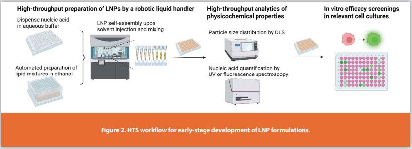
To address the screening needs in early-stage development of LNP formulations, our labs at Genentech Inc. recently developed a HTS workflow, allowing multi-parallel LNP preparation, physicochemical characterizations of particle size distributions and cargo loading, and downstream cellular efficacy assessments, all in 96-well microplate formats (Figure 2).11 Specifically, we used a robotic liquid handler to: 1) dispense the aqueous phase containing the dissolved nucleic acid cargoes in a sample plate; 2) prepare different lipid composition mixtures in ethanol in source plates; and 3) achieve rapid and parallel phase transfer and mixing, which mimicked the solvent-injection process. We optimized multiple injection and mixing process parameters, including speed, phase volume ratio, and phase injection sequence, to reproducibly generate homogeneous LNPs. We also integrated dynamic light scattering (DLS) and UV or fluorescence spectroscopy in the workflow to monitor critical attributes of the resulting formulations prepared in microplates, i.e., particle size distributions and percent nucleic acid loading, respectively. If the majority of screened formulations passed the pre-defined physicochemical characterization criteria, i.e., mono- or poly-dispersed size distributions with mean hydrodynamic diameters less than 200 nm, and encapsulation efficiency around or higher than 80%, the sample plates were then transferred to in vitro efficacy screenings in relevant cell cultures. Compared with the microfluidic formulation method, our HTS approach achieved a formulation scale of 0.1-0.3 mL/micro well, thus saving material inputs by ~10 folds. Furthermore, we were able to screen 96 formulation conditions with analytical readouts of critical physicochemical properties within ~3 h, significantly increasing preparation efficiency by ~100 fold. Being resource-sparing and highly efficient, the HTS workflow helped narrow the screening space in the formulation development cascade of both ASO and mRNA LNPs.
High-Throughput Analytical Characterization of LNPs
According to regulatory guidance, LNP formulations have to be characterized by a panel of physicochemical quality attributes, such as nanoparticle morphology and structure, particle size distribution and concentration, particle surface properties, lipid amount and impurity, unloaded and loaded cargo concentrations, drug release, and stability (Figure 1).12 To be integrated into the HTS workflow, analytics in the microplate format would be preferred. As mentioned earlier, we measured particle size distributions using a DLS plate reader and quantified unloaded and/or total cargo concentrations by using microplate-based spectroscopy (UV or fluorescence), both providing first pass characterization for the screened formulations (Figure 2).

For nucleic acid quantification, the unloaded and loaded portion could be separated by a filtering microplate or size-exclusion column-loaded microplate, which recover the unloaded nucleic acid or loaded LNPs, respectively. Then nucleic acid concentration can be measured by UV intensity at 260 nm or by using appropriate fluorescent probes, such as OliGreen for single-stranded DNA, PicoGreen for double-stranded DNA, and RiboGreen for RNA. These probes can fluoresce upon specific binding with nucleic acids, and can be added to the unpurified samples to only detect the unloaded nucleic acids soluble in the buffer.13 Surfactants are also usually added to disrupt LNPs and extract the loaded nucleic acids, allowing quantification of the loaded and total cargo concentrations. Encapsulation efficiency can then be calculated as the concentration ratio between the loaded and total cargo. Although both the UV and fluorescent probe approaches are compatible with high-throughput measurements, sample pre-processing and preparation of specific external standards are required, limiting seamless in-line analytics. In addition, the measurement accuracy may be affected by the recovery of analytes following purification and/or extraction processes. To better streamline the nucleic acid quantification process in the HTS workflow, recently our labs at Genentech Inc. have investigated UV spectroscopy-based statistical modeling approaches.14 Specifically, the general UV absorbance feature of nucleic acids at ~260 nm as well as their subtle spectral differences between the unloaded and loaded states allowed identification of the unloaded portion from a locally-weighted, partial least square model with inputs of sample UV spectra. A training library was built with spectral data from historical screenings. For each specific unknown sample, a locally weighted regression approach was applied to select appropriate training samples with the most similar spectral features as the unknown sample, so as to form a local training set for concentration prediction. Applied to LNP screenings, this modeling approach predicted cargo loading in similar rank orders as experimental approaches including both UV and fluorescent probe measurements (Figure 3A). Furthermore, it only requires a UV spectrum scan of unpurified samples in microplates, avoiding complicated sample pre-processing and the need of external standards, thus significantly improving the analytical throughput. It also highlights data mining opportunities from HTS of LNPs. Continuous expansion of the training library with more screening samples would further improve the prediction accuracy, thus the model could better serve future LNP screenings. Similarly, Raman spectroscopy-based modeling approaches can be explored to both qualitatively and quantitatively characterize not only nucleic acid cargoes but also lipid components (Figure 3B).15\
Drug release and formulation stability would require kinetic analysis under relevant incubation conditions. For high-throughput analysis, the concentration and chemical stability of released nucleic acids could be analyzed by spectroscopy approaches; while physical stability could be monitored by DLS. In addition to particle size distribution and nucleic acid quantification, microplate-based small-angle X-ray scattering (SAXS) has been recently explored for high-throughput analysis of LNP structures.16 Compared with cryogenic transmission electron microscopy (cryo-TEM), SAXS data provided correlated nanoparticle structure insights while significantly reduced sample preparation efforts owing to automation. In summary, high-throughput DLS, SAXS, and advanced spectroscopy technologies show great promise for seamless integration of quality control processes in the HTS of LNP formulations.
Translation of HTS Results to Scale-Up Formulations
We have compared physicochemical properties of representative ASO LNPs from HTS with the scale-up formulations prepared by microfluidics.11 Same critical formulation variables have been identified with similar impacts on LNP attributes at both formulation scales: higher molar content of the PEGylated lipid incorporated in the lipid composition reduced particle size while increased poly-dispersity, and lower N/P ratio reduced encapsulation efficiency. The two methods also produced ASO LNPs with similar nanoparticle structures as determined by cryo-TEM. Next, we applied the workflow to screen a library of PEGylated lipids for knockdown efficacy in neuron cell cultures.16 Lead formulations identified from HTS were validated by scale-up microfluidic formulations in both physicochemical properties and in vitro efficacy, therefore demonstrating the successful translation of HTS findings. Recently, Cui et al. developed similar screening approaches for mRNA LNPs, also demonstrating successful efficacy translation of LNPs prepared by a robotic liquid handler to the scale-up microfluidic formulations.17 Some HTS formulations showed higher in vitro efficacy than their scale-up counterparts, probably due to a larger particle size and enriched mRNA loading in the particle core as determined by DLS and SAXS, respectively.18 The streamline integration of high-throughput analytics with downstream cellular efficacy readouts facilitates investigations on structure-activity relationships (SAR), and determination of critical formulation design factors and their levels for optimal efficacy. Furthermore, recent development of barcode screening strategies allow HTS of LNPs directly for in vivo efficacy, bridging the in vitro and in vivo translation.19,20

Conclusion
In the emerging era of nucleic acid-based gene therapies and vaccines, efficient delivery systems are indispensable and increasing pharmaceutical development eff orts have been devoted to LNP formulations. HTS workflows that address robotic liquid handler-assisted LNP preparation, high-throughput analytical characterizations, and downstream efficacy screenings can optimize LNPs in a large design space with significant saving in resource and increase in efficiency. HTS results have been successfully translated to scale-up formulations in both in vitro and in vivo assessments. Future development of highly integrated HTS workflows capable of characterizing multi-attributes will allow more data mining and SAR-based formulation design, therefore better guiding early-stage LNP formulation development.
References
- Wei, B., A. Goyon, and K. Zhang, Analysis of therapeutic nucleic acids by capillary electrophoresis. J Pharm Biomed Anal, 2022. 219: p. 114928.
- Chaudhary, N., D. Weissman, and K.A. Whitehead, mRNA vaccines for infectious diseases: principles, delivery and clinical translation. Nat Rev Drug Discov, 2021. 20(11): p. 817-838.
- Hou, X.C., et al., Lipid nanoparticles for mRNA delivery. Nature Reviews Materials, 2021. 6(12): p. 1078-1094.
- Cheng, X. and R.J. Lee, The role of helper lipids in lipid nanoparticles (LNPs) designed for oligonucleotide delivery. Adv Drug Deliv Rev, 2016. 99(Pt A): p. 129-137.
- Suk, J.S., et al., PEGylation as a strategy for improving nanoparticle-based drug and gene delivery. Adv Drug Deliv Rev, 2016. 99(Pt A): p. 28-51.
- Kumar, V., et al., Shielding of Lipid Nanoparticles for siRNA Delivery: Impact on Physicochemical Properties, Cytokine Induction, and Efficacy. Mol Ther Nucleic Acids, 2014. 3: p. e210.
- Eygeris, Y., et al., Deconvoluting Lipid Nanoparticle Structure for Messenger RNA Delivery. Nano Lett, 2020. 20(6): p. 4543-4549.
- Cheng, Q., et al., Selective organ targeting (SORT) nanoparticles for tissue-specific mRNA delivery and CRISPR-Cas gene editing. Nat Nanotechnol, 2020. 15(4): p. 313-320.
- Cullis, P.R. and M.J. Hope, Lipid Nanoparticle Systems for Enabling Gene Therapies. Mol Ther, 2017. 25(7): p. 1467-1475.
- Kulkarni, J.A., et al., Lipid Nanoparticle Technology for Clinical Translation of siRNA Therapeutics. Acc Chem Res, 2019. 52(9): p. 2435-2444.
- Fan, Y., et al., Automated high-throughput preparation and characterization of oligonucleotide-loaded lipid nanoparticles. Int J Pharm, 2021. 599: p. 120392.
- Fan, Y., M. Marioli, and K. Zhang, Analytical characterization of liposomes and other lipid nanoparticles for drug delivery. J Pharm Biomed Anal, 2021. 192: p. 113642.
- Hassett, K.J., et al., Impact of lipid nanoparticle size on mRNA vaccine immunogenicity. J Control Release, 2021. 335: p. 237-246.
- Fan, Y., et al., Spectroscopy-Based Local Modeling Method for High-Throughput Quantification of Nucleic Acid Loading in Lipid Nanoparticles. Anal Chem, 2022. 94(25): p. 9081-9090.
- Kapoor, Y., et al., Flexibility in Drug Product Development: A Perspective. Mol Pharm, 2021. 18(7): p. 2455-2469.
- Sarode, A., et al., Predictive high-throughput screening of PEGylated lipids in oligonucleotide-loaded lipid nanoparticles for neuronal gene silencing. Nanoscale Advances, 2022. 4(9): p. 2107-2123.
- Cui, L.L., et al., Development of a high-throughput platform for screening lipid nanoparticles for mRNA delivery. Nanoscale, 2022. 14(4): p. 1480-1491.
- Cui, L., et al., Mechanistic Studies of an Automated Lipid Nanoparticle Reveal Critical Pharmaceutical Properties Associated with Enhanced mRNA Functional Delivery In Vitro and In Vivo. Small, 2022. 18(9): p. e2105832.
- Sago, C.D., et al., High-throughput in vivo screen of functional mRNA delivery identifies nanoparticles for endothelial cell gene editing. Proc Natl Acad Sci U S A, 2018. 115(42): p. E9944-E9952.
- Hatit, M.Z.C., et al., Species-dependent in vivo mRNA delivery and cellular responses to nanoparticles. Nat Nanotechnol, 2022. 17(3): p. 310-318
Subscribe to our e-Newsletters
Stay up to date with the latest news, articles, and events. Plus, get special
offers from American Pharmaceutical Review delivered to your inbox!
Sign up now!