Johannes Kiefer, University of Bremen, Engineering Thermodynamics and MAPEX Center for Materials and Processes
Abstract
The stability of cytostatic drugs is a critical factor influencing therapeutic efficacy, patient safety, and regulatory compliance. Due to their complex and often reactive chemical structures, cytostatics are prone to degradation via mechanisms like hydrolysis, oxidation, photochemical reactions, and thermal stress. Spectroscopic techniques provide essential molecular-level insight into these processes and complement conventional chromatographic methods. This article summarizes the role of key spectroscopic approaches including vibrational, electronic, and magnetic resonance spectroscopy in the assessment of cytostatic drug stability and degradation, with a focus on industrial pharmaceutical applications.
Cytostatic drugs play a central role in cancer therapy but present significant challenges with respect to chemical stability. Even minor degradation can compromise efficacy or generate toxic impurities, making stability assessment a core requirement throughout drug development and product lifecycle management. Regulatory frameworks require detailed characterization of degradation pathways and products, particularly for cytotoxic compounds.
Cytostatics encompass chemically diverse classes such as alkylating agents, antimetabolites, platinum-based compounds, and targeted small molecules. Their instability arises from functional group reactivity, coordination chemistry, and sensitivity to environmental factors such as light, moisture, temperature, and pH. Common degradation mechanisms include hydrolysis of labile bonds, oxidative pathways, ligand exchange reactions, and photodegradation. These processes are often formulation-dependent and may be influenced by excipients, packaging materials, or storage conditions, necessitating analytical methods capable of resolving subtle chemical changes.
Spectroscopy has emerged as a cornerstone of modern stability studies, offering rapid, non-destructive, and structurally informative analysis. Beyond routine quality control, spectroscopic techniques support mechanistic understanding, formulation optimization, and real-time monitoring in pharmaceutical manufacturing. This paper aims at providing an overview of common spectroscopic tools for studying and monitoring cytostatic drugs.
Vibrational Spectroscopy
Vibrational spectroscopic methods are popular tools in the field of pharmaceutical analysis as they allow molecular fingerprinting to deliver both qualitative and quantitative analysis. Moreover, vibrational spectra carry information about molecular interactions such as hydrogen bonds; hence, they provide insights into mixing behavior and chemical reactions at the molecular level.
Infrared (IR) spectroscopy, particularly FTIR (Fourier-transform IR) with ATR (attenuated total reflection) sampling, is widely used in pharmaceutical laboratories for solid-state and formulation analysis. IR spectra provide direct information on functional groups and are sensitive to chemical transformations such as hydrolysis or oxidation. In stability studies, IR spectroscopy is especially valuable for detecting changes in carbonyl, phosphate, or amide groups and for monitoring solid-state transitions, including polymorphism or amorphization in cytostatic formulations.
IR spectroscopy is a means of absorption spectroscopy. Its scattering counterpart, Raman spectroscopy, complements IR by offering strong performance in aqueous systems and through-container analysis. Its low sensitivity to water makes it particularly suitable for injectable cytostatics and infusion solutions. Raman methods are frequently applied for in situ stability monitoring, spatially resolved analysis of solid dosage forms, and detection of formulation heterogeneity. Confocal Raman microscopy enables localized investigation of degradation processes, supporting root-cause analysis in industrial settings.
UV-Visible Spectroscopy
UV-visible (UV-Vis) spectroscopy remains one of the most widely used analytical techniques for the routine monitoring of cytostatic drug stability, particularly during early development and formulation screening. Many cytostatics such as anthracyclines, alkylating agents, and antimetabolites contain conjugated π-electron systems or aromatic moieties that exhibit characteristic absorbance in the UV or visible range.
From an industrial perspective, UV–Vis spectroscopy offers several advantages: minimal sample preparation, short analysis times, and compatibility with aqueous and organic solvents commonly used in pharmaceutical formulations. These features make it especially attractive for high-throughput stability screening, forced degradation studies, and real-time monitoring during stress testing (e.g., light exposure, pH shifts, or oxidative conditions). Importantly, UV-Vis spectroscopy is well suited for kinetic studies of degradation. Changes in absorbance intensity or wavelength shifts can be tracked over time to derive degradation rate constants and activation energies, providing thermodynamic insight into reaction pathways. In photostability testing, UV-Vis plays a dual role: both as an analytical readout and as a diagnostic tool to assess the overlap between drug absorbance spectra and incident light sources.
Cytostatics encompass chemically diverse classes such as alkylating agents, antimetabolites, platinum-based compounds, and targeted small molecules. Their instability arises from functional group reactivity, coordination chemistry, and sensitivity to environmental factors such as light, moisture, temperature, and pH.
However, UV-Vis spectroscopy has inherent limitations in selectivity. Overlapping spectra of parent compounds and degradation products can complicate interpretation, particularly for complex mixtures. As a result, UV-Vis is most powerful when used as a complementary technique, either coupled with chemometric analysis or integrated into hyphenated systems.
Nuclear Magnetic Resonance (NMR) Spectroscopy
NMR spectroscopy is a cornerstone technique for the in-depth characterization of cytostatic drug stability and degradation mechanisms. Unlike optical spectroscopic methods based on electronic transitions, NMR provides direct structural information at the atomic level, making it indispensable for identifying degradation products and elucidating reaction pathways. In stability studies, one-dimensional (1H, 13C) NMR spectroscopy enables the detection of subtle chemical changes such as hydrolysis, oxidation, isomerization, or ring-opening reactions. Chemical shift changes, signal broadening, and the appearance or disappearance of resonances offer clear indicators of molecular transformation. Two-dimensional NMR techniques further enhance structural assignment, particularly for complex or unexpected degradation products.
For industrial applications, NMR spectroscopy is especially valuable in forced degradation and root-cause analysis, where regulatory expectations require not only detection but also structural identification of impurities. It also plays a critical role in comparability studies and lifecycle management, ensuring that changes in formulation or manufacturing do not alter degradation pathways.
Quantitative NMR adds another layer of relevance, allowing absolute quantification of parent compounds and degradation products without the need for reference standards. This is an important advantage when dealing with novel or transient species.
While NMR spectroscopy is resource-intensive in terms of instrumentation, expertise, and sample quantity, its unparalleled structural insight makes it a strategic tool for late-stage development and regulatory documentation rather than routine quality control.
Hyphenated and Complementary Techniques
Spectroscopic methods are often combined with separation techniques, most notably liquid chromatography (LC). Hyphenated approaches such as LC–UV, LC–MS (mass spectrometry), and LC–NMR represent the current gold standard for comprehensive cytostatic stability analysis. These methods combine the separation power of chromatography with the molecular specificity of spectroscopic detection, addressing the complexity inherent in pharmaceutical degradation studies.
Liquid chromatography coupled with UV detection (LC–UV) remains a workhorse in quality control laboratories due to its robustness and regulatory familiarity. It enables the separation of parent compounds from degradation products, overcoming the selectivity limitations of standalone UV–Vis spectroscopy. When paired with diode-array detection, spectral purity analysis further enhances confidence in peak assignment.
Mass spectrometry-based hyphenated methods (LC–MS, LC–MS/MS) provide unmatched sensitivity and molecular specificity, allowing the detection of low-level impurities well below regulatory thresholds. Accurate mass measurements and fragmentation patterns enable rapid identification of degradation products, even in complex matrices such as injectable formulations or combination therapies. From an industrial standpoint, LC–MS is indispensable for impurity profiling, stability-indicating method development, and compliance with ICH (International Council for Harmonisation) guidelines.
LC–NMR, while less commonly deployed due to cost and complexity, occupies a unique niche in advanced stability studies. It allows structural elucidation of degradation products directly after chromatographic separation, without the need for isolation. This capability is particularly valuable when degradation products are unstable or present at low concentrations.
Conclusion
Spectroscopic techniques are indispensable tools for understanding the stability and degradation of cytostatic drugs. Figure 1 gives an overview of the key techniques and what they are used for in this field. By providing molecular-level insight into chemical transformations, they support robust product design, regulatory compliance, and patient safety. As pharmaceutical products and processes continue to evolve, spectroscopy will remain a central element of stability science in industrial practice.
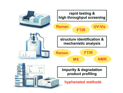
Figure 1. Spectroscopic methods and their application in the study of cytostatic stability and degradation.
The increasing use of spectroscopy in stability studies has driven the adoption of multivariate data analysis. Chemometric tools such as principal component analysis (PCA) and partial least squares (PLS) enable detection of subtle degradation trends and differentiation between normal variability and genuine instability. For industry, these methods support predictive stability modeling, real-time release testing, and implementation of process analytical technology (PAT) frameworks.
Regulatory guidelines require comprehensive understanding of degradation behavior, including identification and qualification of impurities. Spectroscopic techniques provide orthogonal evidence that strengthens stability assessments and enhances method robustness. From an industrial perspective, spectroscopy contributes to faster development timelines, improved product robustness, and reduced risk of late-stage failures. Its compatibility with non-destructive testing and in-process monitoring aligns well with modern pharmaceutical manufacturing strategies.
Ongoing advances in instrumentation, automation, and data analysis are expected to further expand the role of spectroscopy in cytostatic drug development. Integration with continuous manufacturing, PAT, and artificial intelligence-based interpretation will enable more efficient and predictive stability assessments.
References
- International Council for Harmonisation (ICH), Stability Testing of New Drug Substances and Products, ICH Q1A (R2) Scientific Guideline (2003).
- M. Blessy, R.D. Patel, P.N. Prajapati, Y.K. Agrawal, Development of forced degradation and stability indicating studies of drugs-A review, Journal of Pharmaceutical Analysis 4 (2014) 159-165.
- S. Singh, T. Handa, M. Narayanam, A. Sahu, M. Junwal, R.P. Shah, A critical review on the use of modern sophisticated hyphenated tools in the characterization of impurities and degradation products, Journal of Pharmaceutical and Biomedical Analysis 69 (2012) 148-173.
- H. Yilmaz, M. Culha, A Drug Stability Study Using Surface-Enhanced Raman Scattering on Silver Nanoparticles. Applied Sciences 12 (2022) 1807.
About the Author
Johannes Kiefer is professor of engineering thermodynamics at the University of Bremen, Germany and member of the Centre for Process Analytics and Control Technology (CPACT). His research interests include the development of optical spectroscopic methods for engineering and life science applications; in particular, the development of task-specific approaches and multi-parameter measurements including prototype instruments.
Subscribe to our e-Newsletters
Stay up to date with the latest news, articles, and events. Plus, get special
offers from American Pharmaceutical Review delivered to your inbox!
Sign up now!