Introduction
The FDA has adopted the position that all new product submissions for non-sterile drugs must address recovery of Burkholderia cepacia [1,2]. The rationale for this requirement from the review section of the Center for Drug Evaluation and Research (CDER) was published late in 2012 in the trade literature [3]. Both the published article and the regulatory requests have noted the disturbing ability of the Bcc (Burkholderia cepacia complex) group to proliferate in normally well-preserved products and their ability to cause serious complications in susceptible populations [4]. The Agency has expressed concern that “acclimated” Bcc organisms may not be recovered by standard microbiological methods and so evade detection [2]. The potential failure of these methods is of special concern as Bcc organisms have been implicated in a series of FDA recalls for both sterile and non-sterile products. The product types included eyewash, nasal spray, mouthwash, anti-cavity rinse, skin cream, baby and adult washcloths, surgical prep solution, electrolyte solution, and radio-opaque preparations [5]. B. cepacia complex organisms have also been implicated in a series of outbreaks in hospital settings and have earned their reputation as objectionable organisms in specific product categories [6].
This study investigates the concern that compendial methods (especially the use of rich nutrient recovery agar) may not be capable of recovering Bcc microorganisms that had been acclimated to an environment of USP Purified Water under refrigeration (2-8°C) for an extended period of time (up to 42 days). This acclimation method is one suggested specifically for B. cepacia in a pharma environment [2,7].
The Burkholderia cepacia Complex
Members of the Burkholderia cepacia complex are gram-negative bacteria of the β-proteobacteria subdivision and include plant, animal, and human pathogens, with a widespread distribution in natural and man-made habitats [8]. These bacteria exhibit an extraordinary metabolic versatility, allowing their adaptation to a wide range of environments including nutritionally limited ones [9]. Burkholderia cepacia was first described by Burkholder as an agent causing bacterial soft rot in onions [10].
The genus Burkholderia currently comprises more than 60 species. This genus was proposed in 1992 to accommodate the former rRNA group II pseudomonads [10]. Taxonomy of the entire Burkholderia genus has changed rapidly: for instance, B. cepacia has gone from a single species to being a complex comprising 17 closely related species, or genomovars (see Table 1), which can only be correctly classified by using a combination of multiple molecular diagnostic procedures. The literature published prior, and sometimes after, the definition of the cepacia complex identified all the Bcc species as Burkholderia cepacia (or Pseudomonas cepacia), leading to some confusion. Several Bcc strains have developed beneficial interactions with their plant hosts and have proven to be very efficient biocontrol, bioremediation, or plant-growth promoting agents [12-14]. Refer to Table 1 for an overview of the Burkholderia cepacia complex.
Table 1. Overview of the Burkholderia cepacia Complex*

In the past two decades, Burkholderia cepacia has also emerged as an opportunistic human pathogen causing numerous outbreaks, particularly among cystic fibrosis (CF) and other immunocompromised patients. One highly transmissible strain has spread across North America and Britain, and another between hospitalized CF and non-CF patients [15]. In addition, Burkholderia cepacia is inherently resistant to multiple antibiotics and molecular epidemiology, and phylogenetic studies demonstrate that highly transmissible strains emerge randomly; the organism has a capacity for rapid mutation and adaptation (facilitated by numerous insertion sequences) and a large, complex genome divided into separate chromosomes.
An interesting side note on B. cepacia physiology was recently described by Vial et al. [7], who described experiments showing that Bcc can survive and grow within the vacuoles of both amoeba and mammalian macrophages and monocytes. Nasal mucosa has been known to carry amoeba and “consequently could represent an important natural reservoir for Bcc strains and act as a Trojan horse allowing bacteria to access the respiratory tract” [7]. These authors also demonstrated the growth of Bcc in the glucose-rich nutrient medium following the cultivation of the eucaroytic cells. Therefore, this medium type (ATCC Medium 712 PYG) was included in the studies to recover Bcc after growth of amoeba in the medium.
Methodology
Organism preparation
Three distinct types of organisms from the Burkholderia cepacia complex were obtained from the American Type Culture Collection (ATCC a): Burkholderia cepacia (Bc) ATCC 25608, Burkholderia cenocepacia (Bceno) ATCC BAA-245, and Burkholderia multivorans (Bm) ATCC BAA-247 [16]. Organisms were chosen based on discussions with FDA, availability from ATCC, source, and nomenclature history. The organisms were reconstituted as per ATCC instructions, cultured, and then frozen and stored at -70°C using an internal seed lot technique. Two cryovials per organism type were defrosted and transferred onto two trypticase soy agar (TSA) slants for this study. After 48 hours of incubation at 30-35°C, the slants were rinsed with sterile phosphate buffer (SPB) and combined per each organism type. Each slurry of organism was diluted and added to 500mL sterile USP Purified Water to yield 103-104 organisms per mL. Seven bottles were prepared per each organism (one bottle for each time point of testing). Inoculated bottles were kept overnight at room temperature and then transferred into a refrigerator (2-8°C) for the rest of the study to create a lownutrient/ low-temperature environment.
Materials
Both liquid and solid media were employed during this test (see Table 2). The selection of media was based on compendial test methods (USP <61>, Microbiological Examination of Nonsterile Products Microbial Enumeration Tests, and USP <62> Microbiological Examination of Nonsterile Products: Tests for Specified Microorganisms), commonly used environmental test methodology media, and media that were documented to be used for isolation recovery of Burkholderia cepacia.
Table 2. Media Used for Testing (Liquid and Solid)

Acanthamoeba castellanii and Amoeba-enriched medium (Aem)
Acanthamoeba castellanii (AC) ATCC 30234 was reconstituted as per manufacturer instructions and transferred into a 16 × 125mm plastic test tube with 5mL ATCC medium 712 PYG. The culture was incubated at 25°C at approximately 15° horizontal slant. To maintain culture, 0.25mL was transferred into 5mL fresh ATCC medium 712 PYG every 10-11 days of incubation (multiple tubes were created at each transfer). Amoeba- Enriched Medium (AEM) was prepared as follows: AC was grown for 4 days at 25°C in ATCC Medium 712 PYG (ATCC b). After 4 days of incubation AC was removed by centrifugation and the medium was filter-sterilized using a 0.22-micron sterile filter. At this point the medium 712 PYC was denoted as Amoeba-Enriched Medium (AEM). This AEM was then evaluated for its ability to support growth of acclimated Bcc.
Experimental design
Acclimation (Stress) of Organisms in Cold Environment
Organisms were acclimated by storage at 2-8°C for a total of six weeks in Sterile USP Purified Water as described above. Time zero was defined as no more than 24 hours after the inoculated bottles were refrigerated. The organism’s suspensions were tested at weekly intervals marked as day 7, 14, 21, 28, 35, and 42 during the acclimation period. Each interval was tested to determine recoverable organisms by the following methods (for media types refer to the previous section and Table 2):
- Burkholderia cepacia Selective Agar (BCSA) Count: A 1:10 dilution from each acclimation bottle was spread in duplicate on BCSA without supplement for count confirmation.
- Epiflourescence Dye Count: 30mL from each bottle was filtered through 25mm diameter stainless steel filter holders pre-loaded with filter for live/dead epifluorescence test method using a bacterial viability kit. A 95% confidence level was applied to the test results [25,26]. The purpose of this treatment is to provide a culture-independent estimate of the number of dead vs living bacterial cells in the sample.
- Liquid Media Enhancement Comparison: A 1:10 dilution of each acclimation bottle was made into liquid media type (see Table 2 for listing) in duplicate. One set of tubes was placed into a 30-35°C incubator and the second set was placed into a 20-25°C incubator for total of 48 hours. All tubes were then streaked onto each of five solid media types (Cetrimide, TSA, oxidation-fermentation polymyxin bacitracin lactose [OFPBL], and Burkholderia cepacia Selective Agar [BCSA] with and without supplement) after both 24 and 48 hours of incubation (unless growth was confirmed within 24 hours). Streaked plates were incubated at 30-35°C for up to 48 hours. Growth was checked after 24 hours and 48 hours. If growth was observed after 24 hours further testing was discontinued. The liquid media used are listed in in Table 2.
- Membrane Filtration Enhancement Study: 100mL from each inoculated bottle was filtered in triplicate through 0.22-micron (nominal pore size) filters. Filters were placed into 100mL bottles with TSB+LT, R2A Broth, and BCSB that were incubated at 30-35°C for a total of 48 hours. Bottles with filters were streaked onto each of five solid media types (Cetrimide, TSA, OFPBL, and BCSA with and without supplement) after both 24 and 48 hours of incubation (unless growth was confirmed after 24 hours). Streaked plates were incubated at 30-35°C for up to 48 hours.
- Direct Plating: 0.1mL from each inoculated bottle was spread on Total Count Agar Strips using the spread plate method and incubated at 30-35°C for up to 3 days. These strips were used to represent a common media presentation used in environmental monitoring of air.
- R2A Filter Recovery Study: 1mL and 100mL of each organism were filtered through 0.22-micron filter onto R2A Agar and incubated at 30-35°C for up to 5 days. This study was performed to mimic standard water testing using minimal media.
Results
Study #1 (as described above) was performed to count live Bcc microorganisms surviving in a low-nutrient, low-temperature environment (USP Purified Water at 2-8°C) by culturing. The Epiflourescence test methodology (Study #2) was able to enumerate viable microorganisms even if the bacteria were unable to be cultured. Counts obtained by spread plate method on BSCA without addition of supplement (see Figure 1) were compared to counts obtained by epifluorescent live cell count (Figure 2).
 Figure 1.
 Burkholderia cepacia
complex organisms recovery on Burkholderia cepacia
Agar w/o supplements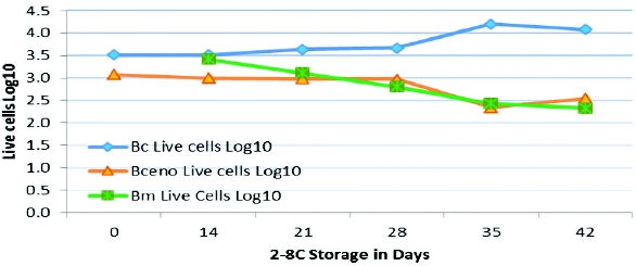 Figure 2. Live Cells Count by Epifluorescent Method for
 Burkholderia cepacia
complex organismsIt was noted by both counting methods that counts for Burkholderia cepacia increased after several weeks at 2-8°C, while counts for Burkholderia cenocepacia and Burkholderia multivorans declined moderately. Epifluorescence counts of live and dead cells were also compared (Figures 2 and 3). There was no direct correlation observed between counts for live cells vs counts for dead cells over time. This might be due to the formation of a biofilm structure or to inherent variability in the test.
 Figure 3. Dead Cells Count by Epifluorescent Method for
 Burkholderia cepacia
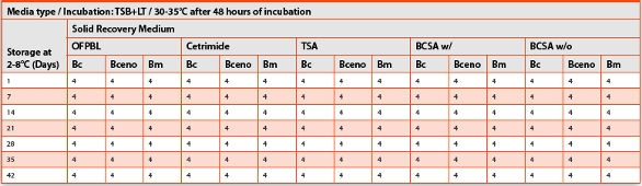
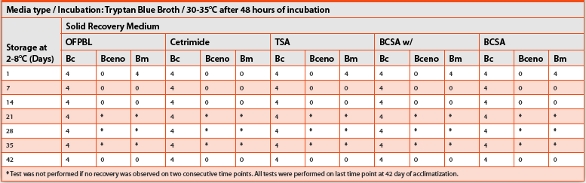
complex organismsStudy #3 evaluated the ability of broth enrichment media to recover Bcc organisms. This was measured by streaking onto solid agar medium (Test #3, see above). This ability was measured semi-quantitatively using the key listed below. The recovery was grouped by medium type and incubation temperature of the broth. Table 3 provides these data where the recovery was recorded as follows: 0 = no recovery, 1 = little growth to no growth, 2 = little growth, 3 = little growth/ sufficient growth, and 4= sufficient growth. Successful recovery was considered when “sufficient growth” was observed on the solid media after 48 hours of enrichment broth incubation and 48 hours of solid media incubation: Bc—Burkholderia cepacia, Bceno—Burkholderia cenocepacia, Bm—Burkholderia multivorans.
Table 3. Evaluation of Microbial Growth from TSB+LT incubated at 30-35°C
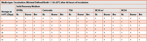
It was of interest in Study #3 that variations were evident between liquid media recovery of Bcc when incubated at different temperatures. An example of these differences is shown in Tables 4 and 5 when the recovery rate is compared between 30-35ºC and 20-25°C incubation of Minimal Defined Broth 1 for 48 hours.
Table 4. Evaluation of Microbial Growth from Minimal Defined Broth incubated at 20-25°C
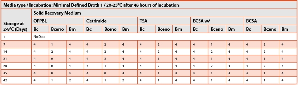
Table 5. Evaluation of Microbial Growth from Minimal Defined Broth incubated at 30-35°C
The least successful liquid media to recover selected organisms was Tryptan Blue Broth; refer to Tables 6 and 7.
Table 6. Evaluation of Microbial Growth from Tryptan Blue Broth incubated at 20-25°C
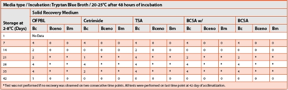
Table 7. Evaluation of Microbial Growth from Tryptan Blue Broth incubated at 30-35°C
It also was observed that through the 42-day acclimation period evaluated, Burkholderia cepacia was the most consistently recovered from all liquid media types after 24 hours of incubation enhancement and plating on all solid agar types with “sufficient growth” (data not shown). The least consistently recovered organism was Burkholderia cenocepacia (see Tables 2 and 3).
All three organisms were resuscitated with “sufficient growth” when 100mL were filtered through 0.2-micron filter and placed into BCB, TSB, and R2A Broth (Test #4).
Acceptable recovery of all three organisms was also observed when 0.1mL was spread over Total Count Agar strips (Test #5) and after filtration of 1mL and 100mL through 0.22-micron filters that were placed on R2A Agar (Test #6).
Discussion/Conclusions
This study was designed to directly address concerns about the ability of standard microbiological test methods to recover cold-water acclimated Bcc organisms. While it is well accepted that the “Absence of Pseudomonas aeruginosa” test in USP is not optimal for recovery of Bcc [27], this study has shown conclusively that acclimated species of Burkholderia cepacia complex (representing different genomovars) could be recovered using a non-specific enrichment step with most compendial media as well as other common media after 48 hours of incubation at 30- 35°C. We also show that although counts of Burkholderia cenocepacia and Burkholderia multivorans were declining throughout the study, they were still detectable by spread plate method and by the epifluorescent live/dead test method. This allows us to conclude that acclimated Burkholderia cepacia complex organisms could be recovered when broth is enriched for 24-48 hours and then streaked for confirmation onto TSA or even more selective media, such as OFPBL or BCSA.
We showed in this study that even very glucose-rich broth (ATCC Medium 712 PYG as well as Amoeba-Enriched Medium) could also successfully recover all three organisms after organisms were acclimated to a low-temperature/low-nutrient environment. However, it should be noted that preparation of these media types is very complex and not necessary for routine testing performed in the quality control microbiology laboratory.
Acknowledgments
The authors want to thank Steve Steinberger and Linda Contiliano of Perritt Laboratories, Inc., for the ordering and preparation of all the media used in the study, and Stephen Carpenter, Ph.D., from Pacific Analytical Laboratory for providing its laboratory support with epifluorescent testing on short notice.
Author Biographies
Julie Barlasov has been the Laboratory Manager at Perritt Laboratories in Hightstown, New Jersey for the past five years. Julie manages a team of microbiologists responsible for testing products, materials, water, and environmental samples for various clients (mostly non-sterile pharmaceutical manufacturers). Julie has an MBA degree in Pharmaceutical Industry from the University of Sciences in Philadelphia and B.S. in Life Science from the Open University of Israel. Julie has been working in the Quality Control Microbiology field for over 14 years. She can be reached at jbarlasov@perrittlab.com.

Scott Sutton has over 25 years of experience in the the pharmaceutical, medical device, cosmetics, and personal products industries with extensive publications and presentations. Consulting and training in GMP, contamination control, investigations of MDD (OOS), laboratory management, and microbiology-related project management are areas of special interest. His clients have included startups, generics, established Fortune 500 companies, law firms, and investment broker houses. Scott has owned and operated The Microbiology Network (http://www.microbiol.org) since 1996. This company provides consulting and training services to industry.

Rick Jakober is the Vice President, Laboratory Services, at Perritt Laboratories, Inc. He earned a B.S. in Environmental Sciences at Cook College, Rutgers University. Mr. Jakober is a seasoned researcher with wide ranging experience in microbiological analyses including in-depth knowledge of FDA and compendial cGMP requirements combined with broad knowledge and experience in USP, Ph. Eur., JP, ISO, ASTM, EPA, and AOAC methodologies. In addition, he has over 30 years’ experience in pharmaceutical microbiology, specializing in non-sterile products. This includes Microbial Content Testing, Antimicrobial Effectiveness Testing, Water Systems, Environmental Monitoring, and Inorganic Chemical Analyses. Mr. Jakober has been with Perritt Laboratories since 1987 and previously was with Carter-Wallace and Ayerst Laboratories. Rick may be reached at rjakober@perrittlab.com.
References
- FDA. 2011 Product Quality Microbiology Review NDA: 202-245 Codeine Sulfate Oral Solution (30 mg/5 mL); http://www.accessdata.fda.gov/drugsatfda_docs/nda/2011/202245Orig1s000MicroR.pdf (accessed 12/19/13).
- J.W. Metcalf. Regulatory Review Perspectives for Non-Sterile Drug Products, 2013. Presented at the 2013 PDA Microbiology Conference in October, 2013.
- L.D. Torbeck, D. Racassi, D. Guilfoyle, R.L. Friesdman, and D. Hussong. Burkholderia cepacia: this decision is overdue. PDA Journal of Pharm. Sci. and Technology 2011; 65(5): 535-543.
- S. Sutton. Letter to the Editor, 2012, in response to L. Torbeck, et al., Burkholderia cepacia: This Decision Is Overdue. PDA Journal of Pharm. Sci. and Technol. 2012, 66(2): 91-95.
- S. Sutton and L. Jimenez. A review of reported recalls involving microbiological control 2004-2011 with emphasis on FDA considerations of “objectionable organisms.” Amer. Pharm. Rev. 2012; 15(1):42-57.
- S. Sutton. What is an objectionable organism? Amer. Pharm. Rev. 2012; 15(6):36-48.
- L. Vial, A. Chapalain, M.C. Groleau, E. Deziel. Minireview–the various lifestyles of the Burkholderia cepacia complex species: a tribute to adaptation. Envir. Microbiol. 2011; 13(1): 1-12.
- E. Mahenthiralingam, T.A. Urban, and J.B. Goldberg. The multifarious, multireplicon Burkholderia cepacia complex. Nature Reviews Microbiol. 2005; 3(2): 144–156.
- W. Beckman and T.G. Lessie. Response of Pseudomonas cepacia to p-lactam antibiotics: utilization of penicillin G as the carbon source. J. Bacteriol. 1979; 140: 1126-1128.
- W. Burkholder. Sour skin, a bacterial rot of onion bulbs. Phytopathology 1950; 40: 115-8.
- L. Chiarini, et al. Burkholderia cepacia complex species: health hazards and biotechnological potential. Trends Microbiol. 2006; 14(6):277-286.
- E. Yabuuchi, Y. Kosako, H. Oyaizu, I. Yano, H. Hotta, Y. Hashimoto, et al. Proposal of Burkholderia gen. nov. and transfer of 7 species of the genus Pseudomonas homology group-II to the new genus, with the type species Burkholderia cepacia. Microbiol. Immunol. 1992; 36(12): 1251–1275.
- J.L. Parke, D. Gurian-Sherman. Diversity of the Burkholderia cepacia complex and implications for risk assessment of biological control strains. Ann. Rev. Phytopathol. 2001; 39: 225-58.
- T. Coenye and P. Vandamme. Diversity and significance of Burkholderia species occupying diverse ecological niches. Environ. Microbiol. 2003; 5(9):719-29.
- A. Holmes, J. Govan, and R. Goldstein. Agricultural use of Burkholderia (Pseudomonas) cepacia: a threat to human health? Emerging Infectious Dis. 1998; 4(2): 221-228.
- ATCC a. Product Sheet for BAA-247 (Burkholderia multivorans), BAA-245 (Burkholderia cenocepacia), and 25416 (Burkholderia cepacia).
- D.J. Reasoner and E.E. Geldreich. A new medium for the enumeration and subculture of bacteria from potable water. Appl. Environ. Microbiol. 1985; 49(1):1-7.
- USP. 2013Microbiological Examination of Nonsterile Products: Microbial Enumeration Tests, andMicrobiological Examination of Nonsterile Products: Tests for Specified Microorganisms. USP36/NF31 vol. 1, The United States Pharmacopeial Convention.
- M.B. Glass, C.A. Beesley, P.P. Wilkins, and A.R. Hoffmaster. Comparison of four selective media for the isolation of Burkholderia mallei and Burkholderia pseudomallei. Am. J. Trop. Med. Hyg. 2009; 80(6): 1023–1028.
- C. Hagedorn, W.D. Gould, T.R. Bardinelli, and D.R. Gustavson. A selective medium for enumeration and recovery of Pseudomonas cepacia biotypes from soil. Appl. Envir. Microbiol. 1987; 53(9): 2265-2268.
- K. Vermis, M. Brachkova, P. Vandamme, and H. Neils. Isolation of Burkholderia cepacia complex genomovars from waters. Systematic and Applied Microbiol. 2003; 26:595- 600.
- ATCC b. Collection of Protists—Production Information Sheet for ATCC 30234 (Acanthamoeba castellanii).
- F.L. Schuster. Cultivation of pathogenic and opportunistic free-living ameobas. Clin. Microbiol. Rev. 2002; 15(3):342-354.
- E.O. King, et al. Two simple media for the demonstration of pyocyanin and fluorescein. J. Lab. Clin. Med. 1954; 44:301-307.
- L. Boulos, L. Live/Dead BacLight: application of a new rapid staining method for direct enumeration of viable and total bacteria in drinking water. J. Microbiol. Meth. 1999; 37:77-86.
- Molecular Probes Invitrogen Detection Technologies, 2004. Live/Dead BacLight Bacterial Viability Kits, Product Information MP07007.
- USP. 1982. Microbial Contamination of Sterile and Non-Sterile Articles, with Special Reference to Pseudomonas cepacia. Pharm. Forum 1982; 8(4):2239.