Abstract
Global regulations that apply to the control of adventitious agents in raw materials cover prions, viruses, mycoplasma, bacteria and molds/yeasts. The establishment of a risk assessment concerning these agents should cover the likelihood of contamination, the consequences if a contamination event occurs and the impact on the product safety and availability to the public. The control of adventitious agents include sourcing of raw materials for prions, testing, cleaning/decontamination, filtration, heat, low pH and gamma irradiation for the remaining agents. The efficacy of these modes of control depends on the agent’s resistance to physical and chemical inactivation and retention by filters. Many viruses may contaminate the biopharmaceutical processes and each family of viruses possess unique resistance to physical and chemical treatments and retention characteristics with filters, thus one approach will not remove or eliminate all viruses. At least ten virus families have been reported to contaminate CHO cells the workhorse for the biopharmaceutical field and the majority of these contaminations have originated from raw materials. A science based approach based on the characteristics of the adventitious agents should be taken to minimize the risk of contamination by prions, viruses, bacteria, mycoplasma and molds/yeasts. Global regulations, current publications and current PDA technical reports aid in the development of this approach.
Global Regulations that Apply to the Control of Adventitious Agents in Raw Materials
Global regulations that give guidance on virus detection that can be applied to raw materials include the USDA 9 CFR 113.53 and 113.47, ICH Q5A, EMA/CHMP/BWP/398498/2005, CBER Guidance for Industry - Cell Substrates and Biomaterials and the Japanese Pharmacopeia 210 [1-6]. Global regulations that give guidance on minimizing the risk of transmitting animal spongiform encephalopathy agents via human and veterinary medicinal products include the EMEA/410/01 Rev.4, and the Japanese Pharmacopeia 210 [7,6]. Regulations that give guidance on the detection of mycoplasma include the USDA 9 CFR 113.28, the European Pharmacopoeia 6.0 Chapter 2.6.7, the Japanese Pharmacopoeia 15, Chapter 14, the FDA “Points to Consider” (1993) and the 21 CFR 610.30 subpart D [8-12].
Adventitious Agent Characteristics
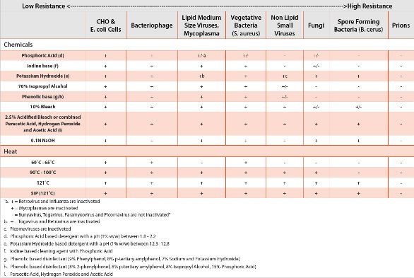
Table 1 - Increasing Resistance to Physical and Chemical Inactivation
Prion proteins exist in at least two forms, the normal or cellular version and the disease causing one. In a process that is not well understood the disease causing prions change normal prions into more disease causing prions. If disease causing prions build up and are not removed by the cell’s machinery, illness can result in the form of spongiform encephalopathy that is fatal. This transmissible spongiform encephalopathy has been reported in deer, elk, cattle, sheep and goats. TSE has also been found in cats that ingested infected bovine material in a zoo setting (Feline SE). TSE in humans is found as an inherited disease (Fatal Familial Insomnia), as a spontaneous occurrence (Creutzfeldt-Jakob Disease (CJD)) or as an infectious disease via ingested infected bovine meat (vCJD). A second route of acquiring vCJD is through a blood transfusion from a vCJD infected donor. An excellent review of prions and the diseases they can cause written by Dr. Stan Prusiner who received the Nobel Prize for his work with prions can be found in the 2004 issue of Scientific America [13].
Table 2. Viruses That Replicate In CHO Cells

Viruses are parasites at the genetic level, thus their replication is strictly dependent on the host’s biochemical machinery. Viruses unlike other adventitious agents must have a cell to replicate in. Viruses are very simple and are made up of a genome that can be either RNA or DNA, a few proteins and some have a lipid envelope acquired from the host. Viruses cannot be viewed by light microscopy and range in size from 200nm to 17nm [14].
Mycoplasma is the trivial name for microorganisms that make up the Mollicutes class of bacteria. These organisms are the smallest free living microorganisms typically about 0.1-0.5 microns in size. They contain no cell wall thus are very pliable and vary in form from round to filamentous. This pliability allows mycoplasma to pass through small pore filtration devices. Since they have no cell wall mycoplasma will not stain in a gram stain.
Table 3 - Source of Virus Contaminations in Raw Materials

Growth requirements for mycoplasma vary. Mycoplasma, in the order Acholeplasma, require limited nutrients allowing them to even replicate in water [15]. A special section in the March 2010 issue of Biologicals showcases nine peer reviewed publications on the nature of mycoplasma and on the new touchdown PCR assay used to detect the mycoplasma to 1 cfu/ml. Also PDA Technical Report No.50 gives an excellent review and consideration for alternative methods for mycoplasma testing [16].
Bacteria are prokaryotic with an absence of a membrane surrounding the nuclear region and no sub cellular organelles. They measure approximately 1 micron and can thrive in a range of environments that is extreme. Bacteria have a cell wall surrounding the cytoplasmic membrane and will thus stain in a Gram stain. Gram positive bacteria like Bacillus and Gram negative bacteria like E. coli are easily identified under a light microscope [17].
Table 4 - Risk Assessment: Adventitious Agents in Raw Materials (Bacteria, Fungi, Mycoplasma and Viruses)
Fungi and molds are eukaryotic containing a membrane bound nucleus and sub cellular organelles. Included in fungi and molds are yeast, filamentous fungi, lichens and slime molds.Their size is approximately 100 microns and is divided into saprobes that feed on dead tissues, mutualists that associate with other species of organisms for mutual aid or parasites that live in or on another living organism with no benefit to the host. Weedy saprobe species are associated with human activities, building and soil are thus of concern in the biopharmaceutical industry [17].
Control of Adventitious Agents in Raw Materials
Control mechanisms include sourcing, testing, cleaning and decontamination of raw material process equipment, filtration, heat, pH and gamma irradiation. The efficacy of these modes of control depends on the adventitious agent’s resistance to physical and chemical inactivation and retention by filters. Generally the least resistant include lipid containing viruses and mycoplasma and the most resistant are bacterial spores followed by Prions as the extreme in resistance [18-20].
Since the process that inactivates prions is extreme (EMEA/410/01 Rev 2) sourcing is a more practical approach for control. A triage approach includes using no raw materials of animal origin, if you must then do not use ruminates (cattle, sheep, goats). If bovine raw materials are used source from a country with negligible BSE risk (e.g. Australia, or New Zealand). Use only animals designated for human consumption and only tissues with no detectable TSE infectivity such as reproductive tissues, musculo-skeletal tissues, trachea, skin or adipose tissue. Also source from animals less than 30 months of age since TSE/BSE is the result of an accumulative of disease causing prions and found generally in older animals. Assure that there is a quality assurance system in place for monitoring the production process and for batch delineation of bovine raw materials. Also self auditing by the suppliers and procedures in place for auditing suppliers should be in place.
Viruses vary in size and makeup that impact their ability to resist inactivation or removal.Many viruses have been reported to replicate in Chinese Hamster Ovary cells (CHO).
Many of these viruses have been reported to have come from contaminated raw materials [21-24].
Some of these raw materials were not of animal origin. If there is a mouse infestation in the storage facility viruses can be transmitted from mice to the outside of raw material container such as salt that can contaminate the raw material when the container is opened. This is especially possible for the small non enveloped viruses in the calicivirus, picornavirus, parvovirus and circovirus families since they are very resistant to inactivation.
Gamma Irradiation is routinely used for the inactivation of medium sized enveloped viruses like Bovine Viral Diarrhea virus (BVDV) in bovine serum [25,26]. The mode of action by gamma irradiation is the disruption of the genome. The general assumption is that the larger the RNA or DNA target the more effective the inactivation. However, in multiple studies the data has shown that multiple factors are responsible for the resistance of viruses to gamma rays [27]. The use of gamma irradiation to inactivate the small viruses in the calicivirus, picorna virus, parvovirus and circovirus families should be done with caution and with a close examination of radiation biology of gamma irradiation and the matrix effect of the medium of suspension or protein concentration.
Mycoplasmas are inactivated by heat, low pH and gamma irradiation (Table 1) [15]. However, recently biofilms have been reported for mycoplasma resulting in resistance to heat as high as 95C (2009 PDA Mycoplasma Workshop in Berlin Germany). Filtration through a 0.1 micron filter if validated for media matrix effects may be effective in removing mycoplasma however it would not be a sterilizing step as it is for bacteria that are ten times larger than mycoplasma. Low pH seems to be the most robust method for inactivating mycoplasma. The growth and long term survival of a Mollicutes, Acholeplasma laidlawii continues to be a challenge to the biopharmaceutical industry. This organism has been detected in animal and plant Tryptic Soy Broth, complex media that had been heated to 95C and filtered, bovine serum, plant and animal peptones and even water [15,28].
Bacteria and molds/fungi are controlled by inactivation and by robust sterile filtration [17]. Generally because of these controls, raw materials do not contribute to bioburden events in the bioreactor.
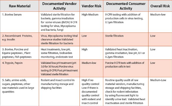
The establishment of a risk assessment concerning these adventitious agents should cover the likelihood of a contamination, the ability to control the agent, the consequences if a contamination event occurs and the impact on the product safety and availability to the public.
The control of these adventitious agents starts with the raw material vendor and ends with the biopharmaceutical user. Some common raw materials are outlined in Table 4 with a risk assessment for the control of the adventitious agents demonstrating that the control of raw materials require a partnership between the vendor and the user.
Some Raw Material Watch Outs
TSE:
Transfection reagents made from lamb’s wool must be sourced from live lambs.Selection antibiotics are often made from bacteria processes that include high risk bovine raw materials (e.g. brain heart infusion broth).
Viruses:
Low pH and gamma irradiation will not remove inactivated viral genome that may be detected by PCR.
Bovine serum may be contaminated with a virus that is not inactivated by gamma irradiation and is not detected in the virus 9 CFR virus testing.
References
- Code of Federal Regulations, title 9 part 113 section53. Requirements for ingredients of animal origin used for production of biologics. US Government Printing Office; 2006. p. 644-645.
- Code of Federal Regulations, title 9 part 113 section 46 and 47. Detection of cytopathogenic and/or hemadsorbing agents and Detection of Extraneous Viruses bt the Fluorescent Antibody Technique.US Government Printing Office; 2006 p. 640-641.
- ICH Q5A: quality of biotechnology products: viral safety evaluation of biotechnology products derived from cell lines of human or animal origin. http://www.ich.org ; 1996.
- EMEA/CHMP?BWP?398498/2005: guideline on virus safety evaluation of biotechnological investigation medicinal products, http://www.emea.europa.eu
- CBER Guidance for Industry. Characterization and qualification of cell substrates and other biological materials used in the production of viral vaccines for infectious disease indications. Office of Communication, Outreach and Development. http://www.fda.gov/BiologicsBloodVaccines/GuidanceComplianceRegulatoryInformation/Guidances/default.htm . 2010
- Society of Japanese Pharmacopoeia 210
- EMEA/410/01 Rev.2, 2004.European medicines agency-Note for guidance on minimizing the risk of transmitting animal spongiform encephalopathu agents via human and veterinary medicinal products.
- Animals and Animal Products, Code of Federal Regulations, Title 9, Part 113; US Printing Office: Washington DC. 2009
- Mycoplasma, general chapter 2.6.7. Ph. Eur.,6th edition. Strasbourg, France: Council of Europe;2007
- Edition XV, Chapter 14. Mycoplasma testing for cell substrates used for the Production of Biotechnological/Biological Products; Society of Japanese Pharmacopoeia: Tokyo, 2006; pp.1721-1724.
- Points to Consider (PTC) in the Characterization of Cell Lines Used to Produce Biologicals; US Food and Drug Administration: Silver Spring, MD, March 1993.
- General Biological Products Standards, Code of Federal Regulations, Title 21, Part 610; US Government Printing Office: Washington DC, 2009.
- Prusiner, S., Scientific American, July 2004.
- Flint, S.J., Enquist, L.W., Racaniello, V.R., Skalka, A.M. editors, The Science of Virology, In Principles of Virology, Molecualr Biology, Pathogenesis and Control of Animal Viruses, ASM Press; Second edition, p2-23, 2004.
- Windsor, H.M., Windsor, DG.D., Noordergraaf, J.H., The growth and long term survival of Acholeplasma laidlawii in media products used in biopharmaceutical manufacturing. Biologicals Volume 38, number 2, March 2010. P204-210.
- PDA Technical Report No.50, Alternative Methods for Mycoplasma testing. 2010. www.pda.org/bookstore .
- Balows, A., Duerden, B.I. editors, Microbiology and Microbial Infections Vol2, Systemic Bacteriology, Oxford University Press, Inc. N.Y., 1998.
- Marsik, F.J., Denys, G.A., Sterilization, Decontamination and Disinfection Procedures for the Microbiology Laboratory. In Murray, P., Baron E.J., Pfaller M.A., Tenover F.C., Yolken R.H., editors. Manual of Clinical Microbiology. Washington D.C.; ASM Press Sixth Edition 1995, p86-98.
- Vesley, D., Lauer, J.L., Decontaminations, Sterilization, Disinfection and Antisepsis. In Fleming, D.O., Richardson, J.H., Tulis, J.J., Vesley, D., editors. Laboratory Safety Principles and Practices. Washington D.C.; ASM Press; Second Edition 1995. Pp219-237.
- Sofer, G., Virus Inactivation in the 1990s and into the 21st Century, Supplement to BioPharm International, June 2003.
- Chen, D., Nims, R., Dusing, S., et.al., Root cause investigation of a viral contamination incident occurred during master cell bank (MCB) testing and characterization- A case study. Biologicals (2008), doi:10.1016/j.biologicals.2008.07.005.
- Fenaux, M., Opriessnig, T., Halbur, P.G., Xu, Y., Potts, B., Meng, X.J., Detection and in vitro and in vivo characterization of porcine circovirus DNA from a porcine-derived commercial pepsin product. Journal of General Virology, vol 85, pp337-3382, 2004.
- Robertson, J.S., Blumel, J., Brorson, K., et.al., Virus & TSE safety forum 2008. Biologicals (2009), doi:10.1016/j.biologicals.2009.05.001.
- Victorial, J.,G., Wang, C., Jones, M., S., et.al., Viral nucleic acids in live-attenuated vaccines: detection of minority variants and adventitious virus. J. Virology. Doi:10.1128/jvi.02690-09 2010.
- Gauvin, G., Nims, R., Gamma-Irradiation of serum for the inactivation of adventitious contaminants. PDA J. Pharm. Sci. Technology. 2010.
- Sullivan, R., Alexander C., Fassolitis, C., et.al., Inactivation of Thirty Viruses by Gamma Radiation. Applied Microbiology vol 22, No1, p61-65, 1971.
- Mahnel, H., Stettmund von Brodorotti, H., Ottis, K., Sensitiveness of viruses for gamma radiation. Zentralbi Bakteriol B. Feb; 170(1-2) p57-70, 1980.
- Kljavin, I., Mycoplasma Contamination in TSB Derived from Plant Peptones. In Proceedings from the PDA Workshop on Mycoplasma Contamination by Plant Peptones. Potts, B.J., editor, pp41-51, 2007’
Author Biography

Dr. Barbara Potts has 28 years experience in the science, compliance and business aspects of the control of adventitious agents (TSE, viruses, mycoplasma) in the bio pharmaceutical, HIV vaccine and immuno therapy, xenotransplantation and cell therapies industries This experience was gained at the University of California, School of Medicine, San Francisco, the NICDS/NIH, NIAID/NIH, University of MN School of Veterinary Medicine, two contract testing companies and three biotechnology companies (Genentech, Repligen and UBI). She is an experimental pathologist with a specialty in virology and has experience from product R&D to IND and BLA acceptance. She has interacted with regulatory agencies, biotechnology companies, Veterinary, Medical, animal owner communities (raw material sources) and recently with the manufacturing diagnostic industry on the development of a commercial PCR kit for the detection of mycoplasma in GMP lot release testing. She holds a B.S. and M.S. in Zoology from Montana State University and a Ph.D. in Experimental Pathology from the University of CA, School of Medicine, San Francisco, CA with an emphasis on viral pathogenesis of the developing CNS.
This article was printed in the March 2011 issue of American Pharmaceutical Review - Volume 14, Issue 2. Copyright rests with the publisher. For more information about American Pharmaceutical Review and to read similar articles, visit www.americanpharmaceuticalreview.com and subscribe for free.